Scientia Agricultura Sinica ›› 2026, Vol. 59 ›› Issue (19): 4400-4416.doi: 10.3864/j.issn.0578-1752.2026.19.017

• ANIMAL SCIENCE·VETERINARY SCIENCE • Previous Articles     Next Articles

Differential Analysis of Hypoxia-Related Factors in the Small Intestines of Yaks and Yellow Cattle

ZHANG YeQiong1(), WANG Jun1, XU MingWei1, HU JiaQi1, LIU ShaSha1, LAN WenLin1, TAN Xiao1, YANG Kun1,2,3()   

  1. 1 College of Life Science and Engineering, Northwest Minzu University, Lanzhou 730000
    2 Gansu Provincial Technology Innovation Center for Animal Cells, Biomedical Research Center, Northwest Minzu University, Lanzhou 730030
    3 Key Laboratory of Bioengineering and Technology, State Ethnic Affairs Commission, Biomedical Research Center, Northwest Minzu University, Lanzhou 730030
  • Received:2025-12-29 Accepted:2026-04-10 Online:2026-10-01 Published:2026-10-10
  • Contact: YANG Kun

Abstract:

【Objective】This study aimed to investigate the differential expression of hypoxia-related factors—Pyruvate dehydrogenase kinase 1 (PDK1), Hypoxia-inducible factor-1α (HIF-1α), Transforming growth factor-β (TGF-β), Vascular endothelial growth factor (VEGF), Mothers against decapentaplegic homolog 2 (Smad2), and Mothers against decapentaplegic homolog 3 (Smad3)—in the small intestinal tissues of yaks, to elucidate the intestinal adaptation mechanisms of yaks to hypoxic environments.【Method】Small intestinal tissues (duodenum, jejunum, and ileum) from plateau yaks and plain yellow cattle were selected as research subjects. Hematoxylin-eosin staining (H.E), periodic acid-Schiff staining (PAS), Masson’s trichrome staining, and immunohistochemical staining were performed. Tissue sections after staining were photographed under a light microscope. Image analysis software was used to measure villus height (V) and crypt depth (C) in H.E-stained sections, as well as the average optical density (AOD) of PAS, Masson, and immunohistochemical staining. The average optical density value was employed to represent positive expression results, thereby investigating the distribution characteristics and expression differences of hypoxia-related factors in different species and intestinal segments.【Result】H.E staining results indicated that the V/C ratio (villus height/crypt depth), an indicator of intestinal digestion, in both yaks and yellow cattle was significantly higher in the duodenum compared to the jejunum and ileum (P<0.05). Furthermore, the duodenal V/C ratio of yaks was significantly higher than that of yellow cattle (P<0.05). PAS staining showed that PAS-positive substances were expressed with significantly higher intensity in the duodenum than in the jejunum and ileum (P<0.05), with strong positive expression observed in the striated border, goblet cells, and Paneth cells. The expression in all intestinal segments of yaks was significantly higher than in yellow cattle (P<0.05). Masson's trichrome staining results revealed significant interspecies and segmental differences in collagen fiber content within the small intestinal tissues of both yaks and yellow cattle (P<0.05). Specifically, the proportion of collagen fibers in the duodenum and jejunum of yellow cattle was significantly higher than in the corresponding segments of yaks (P<0.05). Immunohistochemical analysis detected distinct positive brown-yellow expression of PDK1, HIF-1α, TGF-β, and VEGF in the small intestinal tissues, primarily localized to the striated border, Paneth cells, and goblet cells. Differences in intestinal gland expression were observed between species: weak positive expression was seen in the duodenal and jejunal glands of yellow cattle, while strong positive expression was observed in the corresponding locations in yaks. Tissue segment analysis showed that the expression levels of these factors were significantly higher in the duodenum than in the jejunum (P<0.05). Regarding species differences, the overall expression levels in yak small intestinal tissues were significantly higher than in yellow cattle (P<0.05). However, the expression patterns of Smad2 and Smad3 differed from those of the aforementioned factors. The expressions of Smad2 and Smad3 in the duodenum were significantly lower than those in the ileum and jejunum, respectively (P<0.05). Furthermore, the expression of Smad2/3 in the duodenum and ileum of yaks was significantly lower than in yellow cattle (P<0.05), while in the jejunum of yaks, it was significantly higher than in yellow cattle (P<0.05).【Conclusion】Results from H&E staining, special staining, and immunohistochemical staining revealed significant differences between yaks and yellow cattle in the morphological structure, carbohydrate distribution, and expression of hypoxia-related factors in the small intestine. These findings suggest that yak intestines possess unique structural and molecular adaptive features to hypoxia, and that factors such as PDK1, HIF-1α, and VEGF may play important roles in the intestinal hypoxic adaptation mechanisms of yaks. This study provides fundamental data for research on intestinal adaptation in high-altitude animals and offers a reference for exploring the mechanisms and prevention of hypoxia-related intestinal diseases.

Key words: yak, small intestine, hypoxia, pyruvate dehydrogenase kinase 1 (PDK1), hypoxia inducible factor 1α (HIF-1α), vascular endothelial growth factor (VEGF)

Fig. 1

Small intestinal tissue sections of yak and yellow cattle (H.E staining) A-C, J-L are H.E staining of the duodenum, jejunum and ileum of yellow cattle and yaks in sequence, 200×; D-F and M-O are the H.E staining of the duodenum, jejunum and ileum of yellow cattle and yaks in sequence, 400×; G-I and P-R are H.E staining of the duodenum, jejunum and ileum of yellow cattle and yaks in sequence, 1000×. (IG: Intestinal gland DG: Duodenal gland. GC: Goblet cells. ML: Muscle layer. AC: Absorptive cells. PC: Paneth cells. MM: Muscularis mucosae. BV: Blood vessels. SB: Striated border. S: Submucosa. The same as below"

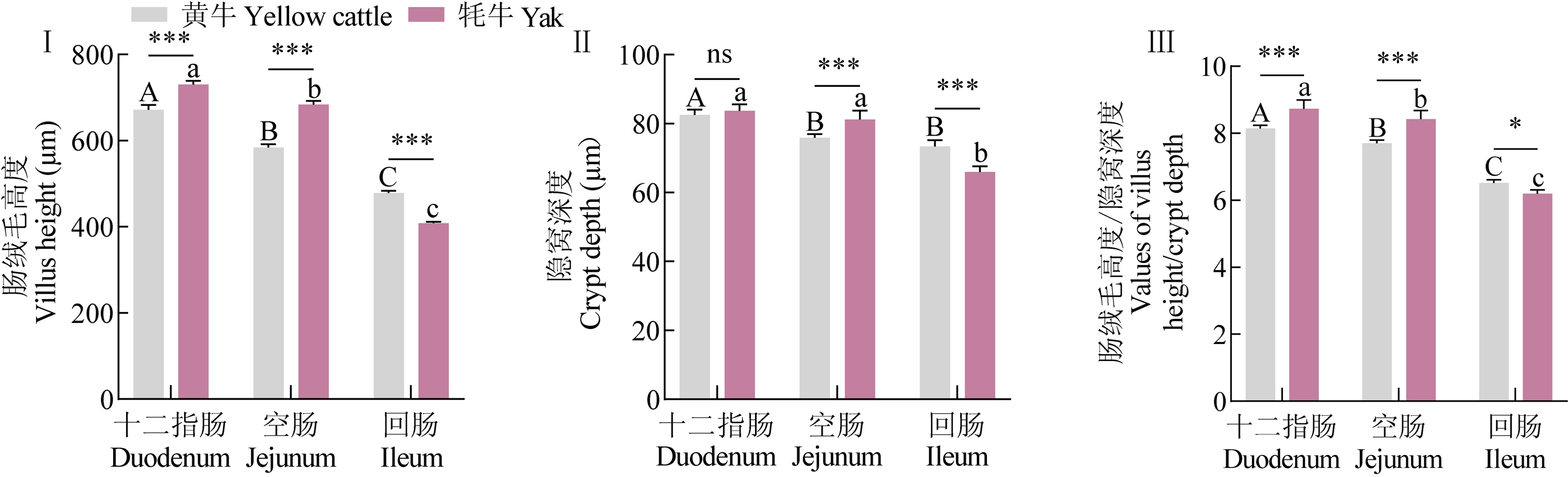
Fig. 2

Morphological measurement of small intestinal tissue in Yaks and yellow cattle (H.E staining) (Ⅰ) Comparison of intestinal villus height among different tissues and species; (Ⅱ) Comparison of crypt depth among different tissues and species; (Ⅲ) Comparison of digestive index (V/C ratio) among different tissues and species. Wherein, ns indicates no significant difference between species (P > 0.05), * indicates a significant difference between species (P < 0.05), ** indicates a very significant difference between species (P < 0.01), and *** indicates an extremely significant difference between species (P < 0.001); different uppercase letters (A, B, C) indicate significant differences among different small intestinal tissues of yellow cattle (P < 0.05), and different lowercase letters (a, b, c) indicate significant differences among different small intestinal tissues of yaks (P < 0.05). The same as below"

Fig. 3

Small intestinal tissue sections of yak and yellow cattle (PAS staining) A-C, J-L are PAS staining of the duodenum, jejunum and ileum of yellow cattle and yaks in sequence, 200×; D-F and M-O are the PAS staining of the duodenum, jejunum and ileum of yellow cattle and yaks in sequence, 400×; G-I and P-R are PAS staining of the duodenum, jejunum and ileum of yellow cattle and yaks in sequence, 1000×"

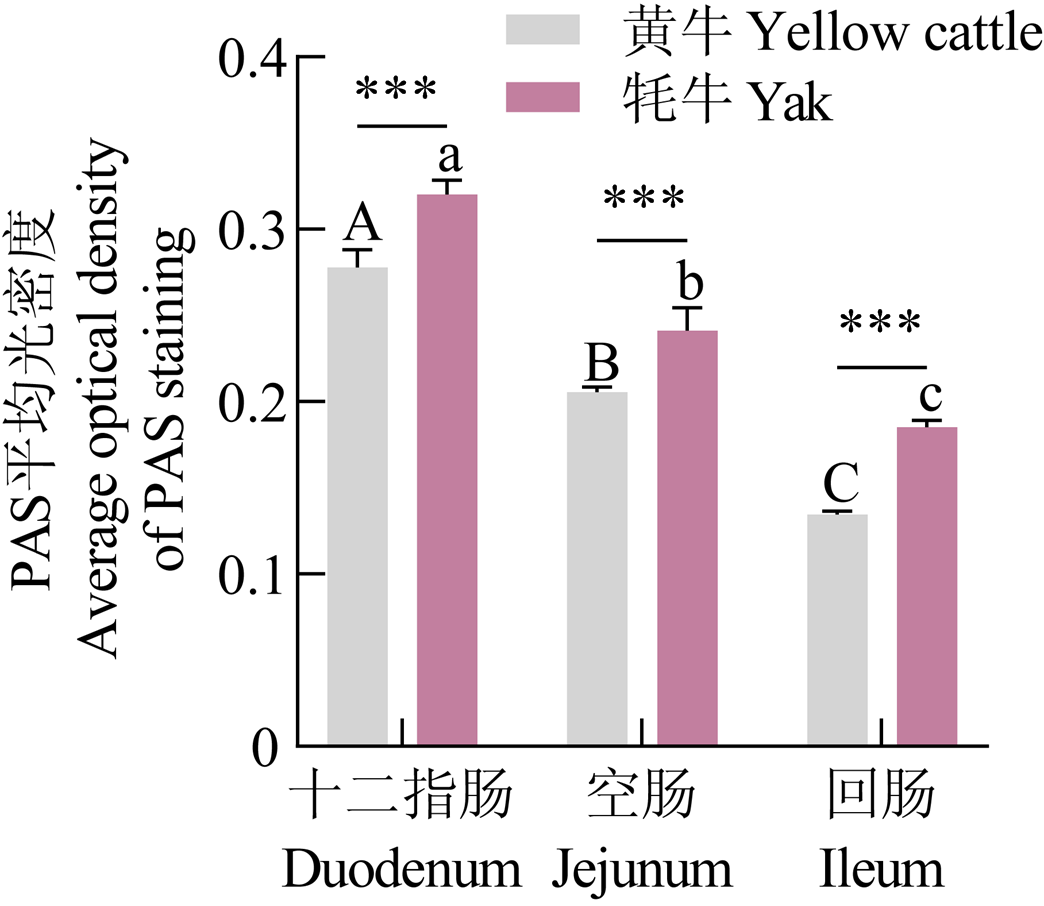
Fig. 4

Morphological measurement of small intestinal tissue in yaks and yellow cattle (PAS staining)"

Fig. 5

Small intestinal tissue sections of yak and yellow cattle (Masson staining) A-C, J-L are Masson staining of the duodenum, jejunum and ileum of yellow cattle and yaks in sequence, 200×; D-F and M-O are the Masson staining of the duodenum, jejunum and ileum of yellow cattle and yaks in sequence, 400×; G-I and P-R are Masson staining of the duodenum, jejunum and ileum of yellow cattle and yaks in sequence, 1000×"

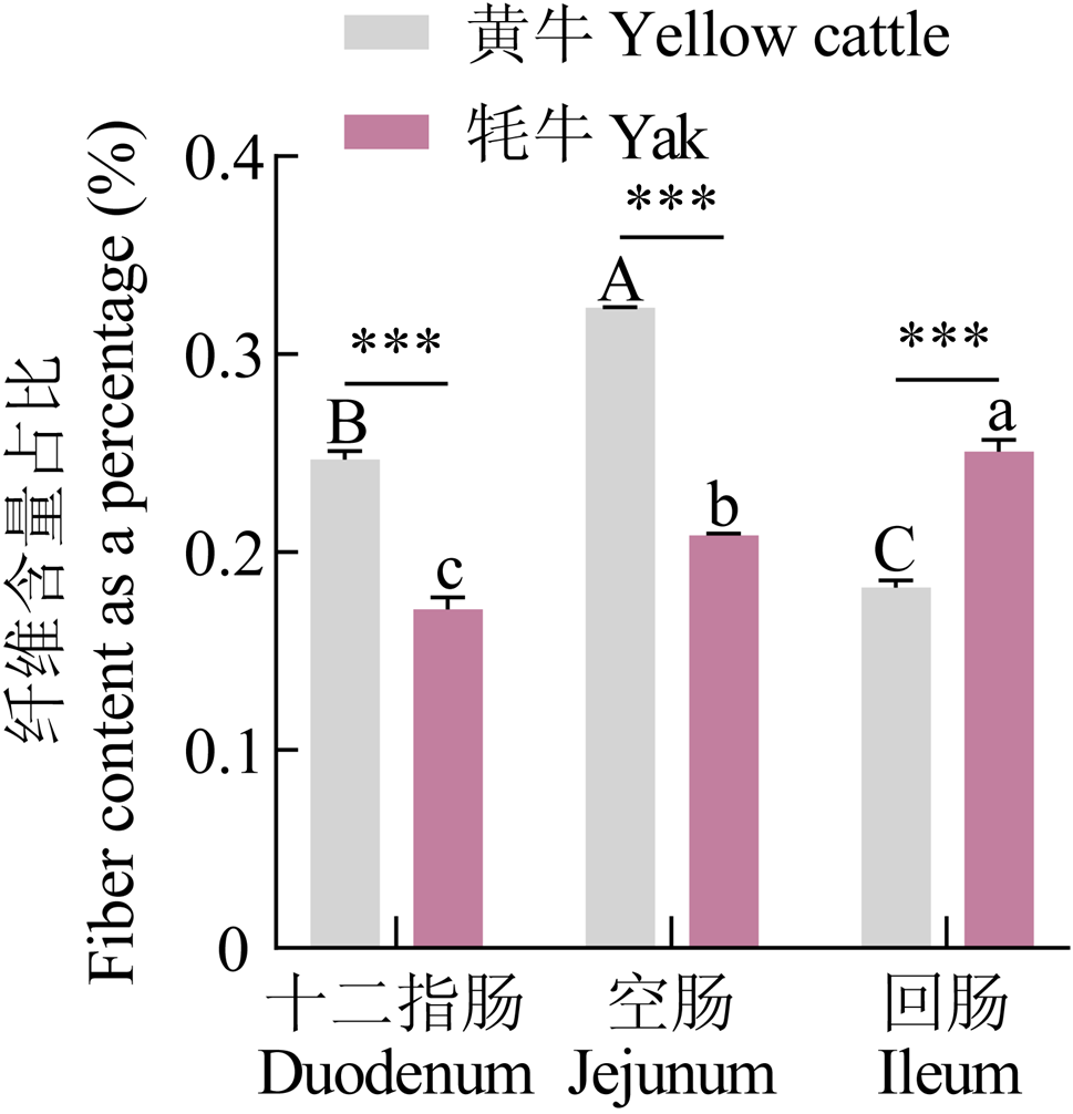
Fig. 6

Morphological measurement of small intestinal tissue in yaks and yellow cattle (Masson staining)"

Fig. 7

Small intestinal tissue sections of yak and yellow cattle (negative control) A-C, J-L are the negative control group of the duodenum, jejunum and ileum of yellow cattle and yaks in sequence, 200×; D-F and M-O are the negative control group of the duodenum, jejunum and ileum of yellow cattle and yaks in sequence, 400×; G-I and P-R are the negative control group staining of the duodenum, jejunum and ileum of yellow cattle and yaks in sequence, 1000×"

Fig. 8

Small intestinal tissue sections of yak and yellow cattle (PDK1 staining) A-C, J-L are PDK1 staining of the duodenum, jejunum and ileum of yellow cattle and yaks in sequence, 200×; D-F and M-O are PDK1 staining of the duodenum, jejunum and ileum of yellow cattle and yaks in sequence, 400×; G-I and P-R are PDK1 staining of the duodenum, jejunum and ileum of yellow cattle and yaks in sequence, 1000×"

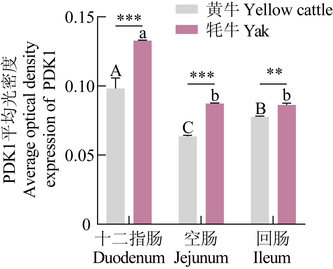
Fig. 9

Morphological measurement of small intestinal tissue in Yaks and yellow cattle (PDK1 staining)"

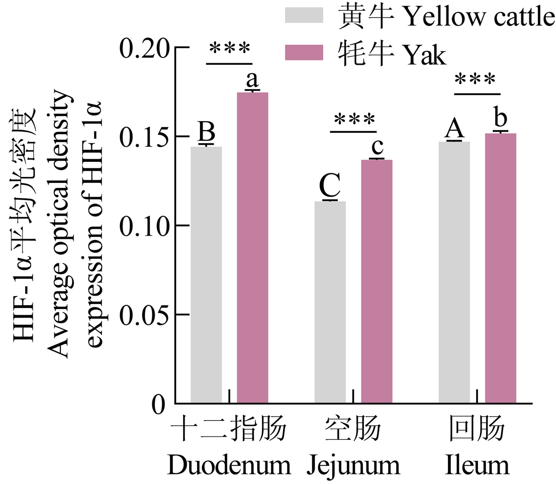
Fig. 10

Small intestinal tissue sections of yak and yellow cattle (HIF-1α staining) A-C, J-L are HIF-1α staining of the duodenum, jejunum and ileum of yellow cattle and yaks in sequence, 200×; D-F and M-O are the HIF-1α staining of the duodenum, jejunum and ileum of yellow cattle and yaks in sequence, 400×; G-I and P-R are HIF-1α staining of the duodenum, jejunum and ileum of yellow cattle and yaks in sequence, 1000×"

Fig. 11

Morphological measurement of small intestinal tissue in Yaks and yellow cattle (HIF-1α staining)"

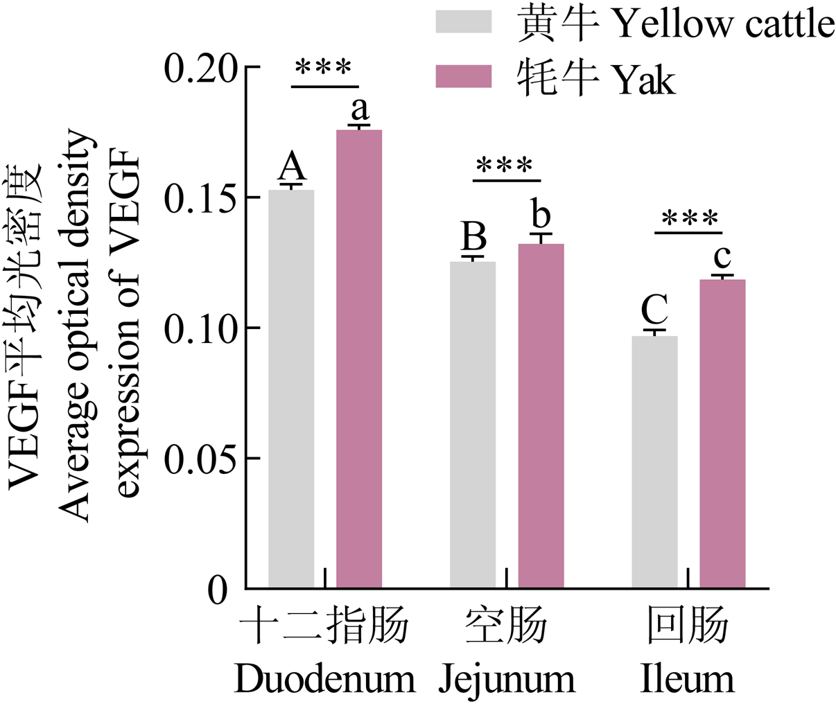
Fig. 12

Small intestinal tissue sections of yak and yellow cattle (VEGF staining) A-C, J-L are VEGF staining of the duodenum, jejunum and ileum of yellow cattle and yaks in sequence, 200×; D-F and M-O were the VEGF staining of the duodenum, jejunum and ileum of yellow cattle and yaks in sequence, 400×; G-I and P-R were VEGF staining of the duodenum, jejunum and ileum of yellow cattle and yaks in sequence, 1000×"

Fig. 13

Morphological measurement of small intestinal tissue in Yaks and yellow cattle (VEGF staining)"

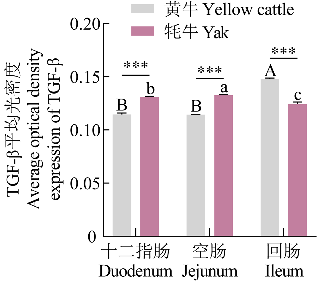
Fig. 14

Small intestinal tissue sections of yak and yellow cattle (TGF-β staining) A-C, J-L are TGF-β staining of the duodenum, jejunum and ileum of yellow cattle and yaks in sequence, 200×; D-F and M-O are the TGF-β staining of the duodenum, jejunum and ileum of yellow cattle and yaks in sequence, 400×; G-I and P-R are TGF-β staining of the duodenum, jejunum and ileum of yellow cattle and yaks in sequence, 1000×"

Fig. 15

Morphological measurement of small intestinal tissue in Yaks and yellow cattle (TGF-β staining)"

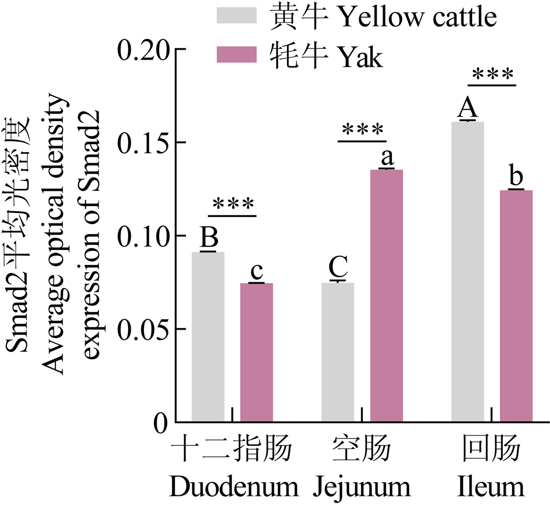
Fig. 16

Small intestinal tissue sections of yak and yellow cattle (Smad2 staining) A-C, J-L are Smad2 staining of the duodenum, jejunum and ileum of yellow cattle and yaks in sequence, 200×; D-F and M-O are the Smad2 staining of the duodenum, jejunum and ileum of yellow cattle and yaks in sequence, 400×; G-I and P-R are Smad2 staining of the duodenum, jejunum and ileum of yellow cattle and yaks in sequence, 1000×"

Fig. 17

Morphological measurement of small intestinal tissue in Yaks and yellow cattle (Smad2 staining)"

Fig. 18

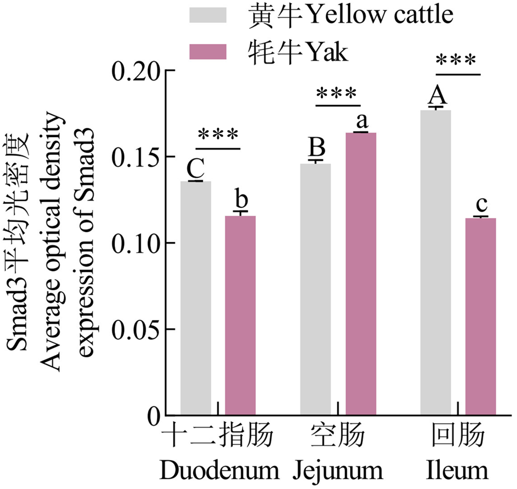
Small intestinal tissue sections of yak and yellow cattle (Smad3 staining) A-C, J-L are Smad3 staining of the duodenum, jejunum and ileum of yellow cattle and yaks in sequence, 200×; D-F and M-O are the Smad3 staining of the duodenum, jejunum and ileum of yellow cattle and yaks in sequence, 400×; G-I and P-R are Smad3 staining of the duodenum, jejunum and ileum of yellow cattle and yaks in sequence, 1000×"

Fig. 19

Morphological measurement of small intestinal tissue in Yaks and yellow cattle (Smad3 staining)"

[1]
杨琦玥, 黄勇, 陈亚冰, 刘洛川, 李键, 兰道亮. 16S rRNA高通量测序技术筛选牦牛瘤胃细菌基因组DNA提取方法及菌群结构[J]. 中国农业科学, 2017, 50(5): 932-941. DOI:10.3864/j.issn.0578-1752.2017.05.016.
Yang Q Y, Huang Y, Chen Y B, Liu L C, Li J, Lan D L. Screening of optimal DNA extraction methods and the bacterial community composition of yak rumen revealed by high 16S rRNA throughput sequencing[J]. Scientia Agricultura Sinica, 2017, 50(5): 932-941. DOI:10.3864/j.issn.0578-1752.2017.05.016. (in Chinese)
[2]
Qiu Q, Zhang G J, Ma T, Qian W B, Wang J Y, Ye Z Q, Cao C C, Hu Q J, Kim J, Larkin D M, Auvil L, Capitanu B, Ma J, Lewin H A, Qian X J, Lang Y S, Zhou R, Wang L Z, Wang K, Xia J Q, et al. The yak genome and adaptation to life at high altitude[J]. Nature Genetics, 2012, 44(8): 946-949.

doi: 10.1038/ng.2343 pmid: 22751099
[3]
Chang E B, Martinez-Guryn K. Small intestinal microbiota: The neglected stepchild needed for fat digestion and absorption[J]. Gut Microbes, 2019, 10(2): 235-240.

doi: 10.1080/19490976.2018.1502539 pmid: 30136893
[4]
Colgan S P, Taylor C T. Hypoxia: An alarm signal during intestinal inflammation[J]. Nature Reviews Gastroenterology & Hepatology, 2010, 7(5): 281-287.
[5]
Taylor C T, Colgan S P. Hypoxia and gastrointestinal disease[J]. Journal of Molecular Medicine, 2007, 85(12): 1295-1300.

doi: 10.1007/s00109-007-0277-z
[6]
Latkowska M, Cai C L, Mitrou M, Marcelino M, Aranda J V, Beharry K D. Gut microbiome and inflammation in response to increasing intermittent hypoxia in the neonatal rat[J]. Pediatric Research, 2025, 97(6): 2126-2135.

doi: 10.1038/s41390-024-03569-7
[7]
Eltzschig H K, Carmeliet P. Hypoxia and inflammation[J]. The New England Journal of Medicine, 2011, 364(7): 656-665.

doi: 10.1056/NEJMra0910283 pmid: 21323543
[8]
Zheng N N, Wei J Q, Wu D P, Xu Y, Guo J P. Master kinase PDK1 in tumorigenesis[J]. Biochimica et Biophysica Acta (BBA) - Reviews on Cancer, 2023, 1878(6): 188971.

doi: 10.1016/j.bbcan.2023.188971
[9]
Roche T E, Baker J C, Yan X, Hiromasa Y, Gong X, Peng T, Dong J, Turkan A, Kasten S A. Distinct regulatory properties of pyruvate dehydrogenase kinase and phosphatase isoforms[J]. Progress in Nucleic Acid Research and Molecular Biology, 2001, 70: 33-75.

pmid: 11642366
[10]
阿依木古丽·阿不都热依木, 张晨, 蔡勇, 覃圣, 罗文学, 扎西英派. 藏绵羊肺脏结构特点及HIF-1α和AQP1的表达特性[J]. 中国农业科学, 2023, 56(11): 2202-2211. DOI:10.3864/j.issn.0578-1752.2023.11.013.
Ayimuguli Abudureyimu, Zhang C, Cai Y, Qin S, Luo W X, Zhaxiyingpai. The micro-structure of Tibetan sheep lung and its HIF-1α and AQP1 expression characteristics[J]. Scientia Agricultura Sinica, 2023, 56(11): 2202-2211. DOI:10.3864/j.issn.0578-1752.2023.11.013. (in Chinese)
[11]
Dolt K S, Mishra M K, Karar J, Baig M A, Ahmed Z, Pasha M A Q. cDNA cloning, gene organization and variant specific expression of HIF-1α in high altitude yak (Bos grunniens)[J]. Gene, 2007, 386(1/2): 73-80.

doi: 10.1016/j.gene.2006.08.004
[12]
Xiong X R, Fu M, Lan D L, Li J, Zi X D, Zhong J C. Yak response to high-altitude hypoxic stress by altering mRNA expression and DNA methylation of hypoxia-inducible factors[J]. Animal Biotechnology, 2015, 26(3): 222-229.

doi: 10.1080/10495398.2014.1002563 pmid: 25927169
[13]
Selak M A, Armour S M, MacKenzie E D, Boulahbel H, Watson D G, Mansfield K D, Pan Y, Simon M C, Thompson C B, Gottlieb E. Succinate links TCA cycle dysfunction to oncogenesis by inhibiting HIF-alpha prolyl hydroxylase[J]. Cancer Cell, 2005, 7(1): 77-85.

doi: 10.1016/j.ccr.2004.11.022 pmid: 15652751
[14]
Wei Y, Zhang D, Shi H, Qian H S, Chen H L, Zeng Q, Jin F F, Ye Y, Ou Z L, Guo M K, Guo B Q, Chen T M. PDK1 promotes breast cancer progression by enhancing the stability and transcriptional activity of HIF-1α[J]. Genes & Diseases, 2024, 11(4): 101041.
[15]
Hou T L, Ma H P, Wang H X, Chen C L, Ye J R, Ahmed A M, Zheng H. Sevoflurane preconditioning attenuates hypoxia/reoxygenation injury of H9c2 cardiomyocytes by activation of the HIF-1/PDK-1 pathway[J]. PeerJ, 2020, 8: e10603.
[16]
Goodwin J, Choi H, Hsieh M H, Neugent M L, Ahn J M, Hayenga H N, Singh P K, Shackelford D B, Lee I K, Shulaev V, Dhar S, Takeda N, Kim J W. Targeting hypoxia-inducible factor-1α/pyruvate dehydrogenase kinase 1 axis by dichloroacetate suppresses bleomycin-induced pulmonary fibrosis[J]. American Journal of Respiratory Cell and Molecular Biology, 2018, 58(2): 216-231.

doi: 10.1165/rcmb.2016-0186OC pmid: 28915065
[17]
Grishko V, Solomon M, Breit J F, Killilea D W, Ledoux S P, Wilson G L, Gillespie M N. Hypoxia promotes oxidative base modifications in the pulmonary artery endothelial cell VEGF gene[J]. FASEB Journal, 2001, 15(7): 1267-1269.

pmid: 11344109
[18]
Liu Y, Cox S R, Morita T, Kourembanas S. Hypoxia regulates vascular endothelial growth factor gene expression in endothelial cells. Identification of a 5' enhancer[J]. Circulation Research, 1995, 77(3): 638-643.

doi: 10.1161/01.res.77.3.638 pmid: 7641334
[19]
Ferrara N, Gerber H P, LeCouter J. The biology of VEGF and its receptors[J]. Nature Medicine, 2003, 9(6): 669-676.

doi: 10.1038/nm0603-669 pmid: 12778165
[20]
de Bock K, Georgiadou M, Carmeliet P. Role of endothelial cell metabolism in vessel sprouting[J]. Cell Metabolism, 2013, 18(5): 634-647.

doi: 10.1016/j.cmet.2013.08.001 pmid: 23973331
[21]
Sharmin N, Nganwuchu C C, Nasim M T. Targeting the TGF-β signaling pathway for resolution of pulmonary arterial hypertension[J]. Trends in Pharmacological Sciences, 2021, 42(7): 510-513.

doi: 10.1016/j.tips.2021.04.002 pmid: 33966900
[22]
林凯, 虞德兵, 解晓东, 于敏莉, 李东锋, 杜文兴. TGF-β/Smad信号通路在Follistatin调节鸭骨骼肌卫星细胞增殖过程中的作用机制[J]. 中国农业科学, 2015, 48(12): 2460-2468. DOI: 10.3864/j.issn.0578-1752.2015.12.019.
Lin K, Yu D B, Xie X D, Yu M L, Li D F, Du W X. The regulated mechanism of follistatin on the proliferation process of duck skeletal muscle satellite cell involved in TGF-β/smad signaling pathway[J]. Scientia Agricultura Sinica, 2015, 48(12): 2460-2468. DOI: 10.3864/j.issn.0578-1752.2015.12.019. (in Chinese)
[23]
郭泽媛, 杜张胜, 张雅琦, 陈春路, 马晓燕, 成颖, 王锴, 吕丽华. Smad7介导TGF-β信号通路对绵羊卵泡颗粒细胞增殖的影响[J]. 中国农业科学, 2023, 56(13): 2597-2608. DOI: 10.3864/j.issn.0578-1752.2023.13.013.
Guo Z Y, Du Z S, Zhang Y Q, Chen C L, Ma X Y, Cheng Y, Wang K, Lü L H. Effects of Smad7-mediated TGF-β signaling pathway on proliferation of sheep granulosa cells[J]. Scientia Agricultura Sinica, 2023, 56(13): 2597-2608. DOI: 10.3864/j.issn.0578-1752.2023. 13.013. (in Chinese)
[24]
Choi Y K. Detrimental roles of hypoxia-inducible factor-1α in severe hypoxic brain diseases[J]. International Journal of Molecular Sciences, 2024, 25(8): 4465.

doi: 10.3390/ijms25084465
[25]
张瑞莉, 葛铭, 郑世民. 缺氧缺血致脑损伤机理的研究[J]. 东北农业大学学报, 2005, 36(6): 774-777.
Zhang R L, Ge M, Zheng S M. Study on pathogenesis of hypoxic- ischemic on brain damage[J]. Journal of Northeast Agricultural University, 2005, 36(6): 774-777. (in Chinese)
[26]
徐一帆, 周陈晨, 刘希成, 郭维华, 周建. 缺氧相关疾病动物模型研究进展[J]. 四川大学学报(医学版), 2025, 56(2): 331-338.
Xu Y F, Zhou C C, Liu X C, Guo W H, Zhou J. Application of animal models in research on hypoxia-related diseases[J]. Journal of Sichuan University (Medical Sciences), 2025, 56(2): 331-338. (in Chinese)
[27]
Nabuurs M J A, Hoogendoorn A, Van Der Molen E J, Van Osta A L M. Villus height and crypt depth in weaned and unweaned pigs, reared under various circumstances in the Netherlands[J]. Research in Veterinary Science, 1993, 55(1): 78-84.

pmid: 8378616
[28]
巨晓军, 章明, 刘一帆, 姬改革, 单艳菊, 屠云洁, 邹剑敏, 张海涛, 卞良永, 束婧婷. 整合肠道菌群与小分子代谢物分析调控肉鸡饲料转化率的影响因素[J]. 中国农业科学, 2025, 58(6): 1223-1238. DOI: 10.3864/j.issn.0578-1752.2025.06.013.
Ju X J, Zhang M, Liu Y F, Ji G G, Shan Y J, Tu Y J, Zou J M, Zhang H T, Bian L Y, Shu J T. Integration of intestinal flora and small molecule metabolite to analyze the role of factors regulating feed conversion in broiler chickens[J]. Scientia Agricultura Sinica, 2025, 58(6): 1223-1238. DOI: 10.3864/j.issn.0578-1752.2025.06.013. (in Chinese)
[29]
Steinbach E, Belda E, Alili R, Adriouch S, Dauriat C J G, Donatelli G, Dumont J L, Pacini F, Tuszynski T, Pelloux V, Jacques F, Creusot L, Coles E, Taillandier P, Vazquez Gomez M, Masi D, Mateo V, André S, Kordahi M, Rouault C, et al. Comparative analysis of the duodenojejunal microbiome with the oral and fecal microbiomes reveals its stronger association with obesity and nutrition[J]. Gut Microbes, 2024, 16(1): 2405547.

doi: 10.1080/19490976.2024.2405547
[30]
王文婷, 邬应龙. 氧化魔芋葡甘露聚糖对高脂饮食C57BL/6J小鼠十二指肠形态及肠道菌群的影响[J]. 食品科学, 2013, 34(19): 306-310.

doi: 10.7506/spkx1002-6630-201319063
Wang W T, Wu Y L. Effect of oxidized konjac glucomannan on duodenal morphology and intestinal flora in C57BL/6J mice fed high-fat diet[J]. Food Science, 2013, 34(19): 306-310. (in Chinese)

doi: 10.1111/jfds.1969.34.issue-4
[31]
李蕊彤, 陈清梅, 徐建芳, 张军民, 司玮, 张铁鹰. 基于猪肠道类器官模型的甘草次酸抗氧化功能及机制研究[J]. 中国农业科学, 2025, 58(22): 4771-4785. DOI: 10.3864/j.issn.0578-1752.2025.22.016.
Li R T, Chen Q M, Xu J F, Zhang J M, Si W, Zhang T Y. Study on the antioxidant function and mechanism of 18β-glycyrrhetinic acid using porcine intestinal organoid model[J]. Scientia Agricultura Sinica, 2025, 58(22): 4771-4785. DOI: 10.3864/j.issn.0578-1752.2025.22.016. (in Chinese)
[32]
Pelaseyed T, Bergström J H, Gustafsson J K, Ermund A, Birchenough G M, Schütte A, van der Post S, Svensson F, Rodríguez-Piñeiro A M, Nyström E E, Wising C, Johansson M E, Hansson G C. The mucus and mucins of the goblet cells and enterocytes provide the first defense line of the gastrointestinal tract and interact with the immune system[J]. Immunological Reviews, 2014, 260(1): 8-20.

doi: 10.1111/imr.12182 pmid: 24942678
[33]
Hansson G C. Mucins and the microbiome[J]. Annual Review of Biochemistry, 2020, 89: 769-793.

doi: 10.1146/annurev-biochem-011520-105053 pmid: 32243763
[34]
Storkholm J H, Villadsen G E, Jensen S L, Gregersen H. Mechanical properties and collagen content differ between isolated guinea pig duodenum, jejunum, and distal ileum[J]. Digestive Diseases and Sciences, 1998, 43(9): 2034-2041.

pmid: 9753270
[35]
Van Soest B, Weber Nielsen M, Moeser A J, Abuelo A, VandeHaar M J. Transition milk stimulates intestinal development of neonatal Holstein calves[J]. Journal of Dairy Science, 2022, 105(8): 7011-7022.

doi: 10.3168/jds.2021-21723 pmid: 35691749
[36]
Zeng Y J, Qiao A K, Yu J D, Zhao J B, Liao D H, Xu X H, Hans G. Collagen fiber angle in the submucosa of small intestine and its application in Gastroenterology[J]. World Journal of Gastroenterology, 2003, 9(4): 804-807.

doi: 10.3748/wjg.v9.i4.804
[37]
Rousset M. The human colon carcinoma cell lines HT-29 and Caco-2: Two in vitro models for the study of intestinal differentiation[J]. Biochimie, 1986, 68(9): 1035-1040.

pmid: 3096381
[38]
Majmundar A J, Wong W J, Simon M C. Hypoxia-inducible factors and the response to hypoxic stress[J]. Molecular Cell, 2010, 40(2): 294-309.

doi: 10.1016/j.molcel.2010.09.022 pmid: 20965423
[39]
Kim J W, Tchernyshyov I, Semenza G L, Dang C V. HIF-1-mediated expression of pyruvate dehydrogenase kinase: A metabolic switch required for cellular adaptation to hypoxia[J]. Cell Metabolism, 2006, 3(3): 177-185.

doi: 10.1016/j.cmet.2006.02.002
[40]
Floor E, Su J Y, Chatterjee M, Kuipers E S, IJssennagger N, Heidari F, Giordano L, Wubbolts R W, Mihăilă S M, Stapels D A C, Vercoulen Y, Strijbis K. Development of a Caco-2-based intestinal mucosal model to study intestinal barrier properties and bacteria-mucus interactions[J]. Gut Microbes, 2025, 17(1): 2434685.

doi: 10.1080/19490976.2024.2434685
[41]
Derynck R, Budi E H. Specificity, versatility, and control of TGF-β family signaling[J]. Science Signaling, 2019, 12(570): eaav5183.

doi: 10.1126/scisignal.aav5183
[42]
Bernard K, Logsdon N J, Ravi S, Xie N, Persons B P, Rangarajan S, Zmijewski J W, Mitra K, Liu G, Darley-Usmar V M, Thannickal V J. Metabolic reprogramming is required for myofibroblast contractility and differentiation[J]. Journal of Biological Chemistry, 2015, 290(42): 25427-25438.

doi: 10.1074/jbc.M115.646984 pmid: 26318453
[43]
McMahon S, Charbonneau M, Grandmont S, Richard D E, Dubois C M. Transforming growth factor beta1 induces hypoxia-inducible factor-1 stabilization through selective inhibition of PHD2 expression[J]. The Journal of Biological Chemistry, 2006, 281(34): 24171-24181.

doi: 10.1074/jbc.M604507200
[44]
Tsukada Y I, Fang J, Erdjument-Bromage H, Warren M E, Borchers C H, Tempst P, Zhang Y. Histone demethylation by a family of JmjC domain-containing proteins[J]. Nature, 2006, 439(7078): 811-816.

doi: 10.1038/nature04433
[45]
Cai L, Sutter B M, Li B, Tu B P. Acetyl-CoA induces cell growth and proliferation by promoting the acetylation of histones at growth genes[J]. Molecular Cell, 2011, 42(4): 426-437.

doi: 10.1016/j.molcel.2011.05.004 pmid: 21596309
[46]
Liu R M, Desai L P. Reciprocal regulation of TGF-β and reactive oxygen species: A perverse cycle for fibrosis[J]. Redox Biology, 2015, 6: 565-577.

doi: 10.1016/j.redox.2015.09.009
[47]
Bai Y H, Huang L L, Fan Y, Li Y L. Marrow mesenchymal stem cell mediates diabetic nephropathy progression via modulation of Smad2/3/WTAP/M6A/ENO1 axis[J]. FASEB Journal, 2024, 38(11): e23729.

doi: 10.1096/fsb2.v38.11
[1] GERIQIMUGE, PUBUZHANDUI, XU Qing, HOU LingLing. Effects of Hypoxia on Proliferation of Bovine Renal Cells and Mitochondrial Autophagy [J]. Scientia Agricultura Sinica, 2026, 59(6): 1333-1347.
[2] LIU ZhuoLin, LIU HongYun. The Potential and Mechanisms of Apigenin to Relieve Heat Stress and Hypoxia in Dairy Cows Based on Network Pharmacology and Molecular Docking [J]. Scientia Agricultura Sinica, 2024, 57(5): 1010-1022.
[3] MENG ZhaoYi, WANG YunLu, YAO YiLong, XI GuangYin, NIU JiaQiang, SOLANGSIZHU, GUO Min, XU YeFen. lncRNA-MSTRG.7889.1 Competitively Binds to bta-miR-146a Targeting Smad4 to Regulate Apoptosis of Yak Granulosa Cells [J]. Scientia Agricultura Sinica, 2024, 57(4): 797-809.
[4] ZHANG Peng, WANG MingXiu, JING KeMin, LI YuQian, TIAN Yuan, ZHONG JinCheng, CAI Xin. Cloning of PLZF Gene and Its Effects on the Proliferation of Undifferentiated Spermatogonia in Cattleyak [J]. Scientia Agricultura Sinica, 2024, 57(2): 390-402.
[5] PAN YangYang, WANG JingLei, WANG Meng, WANG LiBin, ZHANG Qian, CHEN Rui, ZHANG TianTian, CUI Yan, XU GengQuan, FAN JiangFeng, YU SiJiu. Formation and Function of Paraspeckle During Pre-implantation Embryos Development in Yak [J]. Scientia Agricultura Sinica, 2023, 56(6): 1189-1203.
[6] LI JiRong, LIU Xin, WANG Jun, CAO XiaoGang, CI Dun. Fractionation Effect of Stable Carbon and Nitrogen Isotope Ratios in Yak Dairy Products Processing [J]. Scientia Agricultura Sinica, 2023, 56(10): 1982-1993.
[7] WANG JiaMin,SHI JiaChen,MA FangFang,CAI Yong,QIAO ZiLin. Effects of Soy Isoflavones on the Proliferation and Apoptosis of Yak Ovarian Granulosa Cells [J]. Scientia Agricultura Sinica, 2022, 55(8): 1667-1675.
[8] RAN HongBiao,ZHAO LiLing,WANG Hui,CHAI ZhiXin,WANG JiKun,WANG JiaBo,WU ZhiJuan,ZHONG JinCheng. Effects of lncFAM200B on the Lipid Deposition in Intramuscular Preadipocytes of Yak [J]. Scientia Agricultura Sinica, 2022, 55(13): 2654-2666.
[9] CHEN LuLu,WANG Hui,WANG JiKun,WANG JiaBo,CHAI ZhiXin,CHEN ZhiHua,ZHONG JinCheng. Comparative Analysis of miRNA Expression Profiles in the Hearts of Tibetan Cattle and Xuanhan Cattle [J]. Scientia Agricultura Sinica, 2020, 53(8): 1677-1687.
[10] LI XueRu,SHI XiXiong,WANG JianZhong,ZHANG PanGao,TIAN Zhu,HAN Ling. Effect of Nitric Oxide Synthetase Inhibitor on Yak Meat Quality During Post-Mortem Aging [J]. Scientia Agricultura Sinica, 2020, 53(8): 1617-1626.
[11] MIAO JianJun,PENG ZhongLi,GAO YanHua,BAI Xue,XIE XinTing. Effects of Dietary Small Peptides on Production Performance and Expression of PepT1 mRNA in Digestive Tract of Fattening Yaks [J]. Scientia Agricultura Sinica, 2020, 53(23): 4950-4960.
[12] PAN YangYang,WANG Meng,RUI Xian,WANG LiBin,HE HongHong,WANG JingLei,MA Rui,XU GengQuan,CUI Yan,FAN JiangFeng,YU SiJiu. RNA-Binding Motif Protein 3(RBM3) Expression is Regulated by Insulin-Like Growth Factor (IGF-1) for Protecting Yak (Bos grunniens) Cumulus Cells from Apoptosis During Hypothermia Stress [J]. Scientia Agricultura Sinica, 2020, 53(11): 2285-2296.
[13] SUN Kai, LI DongXiu, YANG Jing, DONG JiChi, YAN XianCheng, LUO LiXin, LIU YongZhu, XIAO WuMing, WANG Hui, CHEN ZhiQiang, GUO Tao. Genome-Wide Association Analysis for Rice Submergence Seedling Rate [J]. Scientia Agricultura Sinica, 2019, 52(3): 385-398.
[14] XiangYue HUANG,XianRong XIONG,Jie HAN,XianYing YANG,Yan WANG,Bin WANG,Jian LI. Expression Pattern of KDM1A in the Development of Yak Follicles [J]. Scientia Agricultura Sinica, 2019, 52(24): 4624-4631.
[15] ZI XiangDong, LUO Bin, XIA Wei, ZHENG YuCai, XIONG XianRong, LI Jian, ZHONG JinCheng, ZHU JiangJiang, ZHANG ZhengFan. Transcriptomic Analysis of IVF Embryonic Development in the Yak (Bos grunniens) Via RNA-Seq [J]. Scientia Agricultura Sinica, 2018, 51(8): 1577-1589.
Viewed
Full text


Abstract

Cited

  Shared   
  Discussed   
No Suggested Reading articles found!