Scientia Agricultura Sinica ›› 2026, Vol. 59 ›› Issue (14): 3220-3236.doi: 10.3864/j.issn.0578-1752.2026.14.016

• ANIMAL SCIENCE·VETERINARY SCIENCE • Previous Articles    

MOTS-C Regulates Mitochondrial Dynamics and Oxidative Stress to Inhibit Apoptosis and Enhance Energy Metabolism in IPEC-J2 Cells

HUO LeLe(), TIAN WanRu, WANG BinBin, CAO HanYang, LI MengXuan, LIU JunYing, LUO Gang, SHEN ManMan, SUN LiuMei, LIU JiYing()   

  1. Jiangsu Key Laboratory of Sericultural and Animal Biotechnology/School of Biotechnology, Jiangsu University of Science and Technology/Key Laboratory of Silkworm and Mulberry Genetic Improvement, Ministry of Agriculture and Rural Affairs/Sericultural Scientific Research Center, Chinese Academy of Agricultural Sciences, Zhenjiang 212100, Jiangsu
  • Received:2025-11-29 Accepted:2026-05-07 Online:2026-07-16 Published:2026-07-21
  • Contact: LIU JiYing

Abstract:

【Objective】By obtaining the genetic sequence of MOTS-C and analyzing its effects on mitochondrial dynamics and antioxidant capacity in porcine intestinal epithelial cells (IPEC-J2), its effects on cell proliferation and intestinal barrier were detected, so as to provide a new molecular target and theoretical basis for the use of MOTS-C to improve piglet intestinal health.【Method】Overexpression vectors or exogenous MOTS-C peptides were used to treat IPEC-J2 cells, and their effects on cell proliferation, barrier function, mitochondrial dynamics, oxidative stress, and apoptosis were evaluated.【Result】Pig MOTS-C was encoded by a highly conserved 21-amino acid peptide from the mitochondrial genome. In IPEC-J2 cells, MOTS-C overexpression significantly downregulated the mitochondrial dynamics-related genes mRNA levels of DRP1 (P<0.01) and Fis1 (P<0.05), while upregulating the fusion-promoting protein OPA1 (P<0.01) and the protein expression of MFN2 (P<0.01). MOTS-C reduced the production of ROS, increased the activities of CAT and T-SOD (P<0.01), lowered MDA content (P<0.01), and upregulated antioxidative genes, including superoxide SOD3 and CAT (P<0.05) at the mRNA level, as well as SOD2 and GCLM at the protein level (P<0.05). MOTS-C also enhanced the expression of tight junction-related genes, including Claudin and Occludin (P<0.05), and proliferation-associated genes, such as CDK2 and CCNDBP1 (P<0.05), and increased CCND1 protein expression (P<0.05). In addition, MOTS-C suppressed the mRNA expression of pro-apoptotic genes BAX and CASP3 (P<0.05), while elevating the protein level of the anti-apoptotic gene BCL2 (P<0.05). MOTS-C peptide significantly downregulated the mRNA levels of DRP1 (P<0.05) and FIS1 (P<0.01), while significantly upregulating OPA1 (P<0.05). Western blot results showed that the expression of MFN2 protein was increased (P>0.05). MOTS-C peptide also significantly increased CAT enzyme activity levels (P<0.05) and significantly reduced intracellular MDA content (P<0.05). Moreover, it significantly increased SOD3 and HO-1 mRNA levels (P<0.05). Further Western blot analysis indicated MOTS-C peptide significantly increased HO-1 protein level, and it consistent with the trend in mRNA levels (P<0.05). In addition, the MOTS-C peptide downregulated the mRNA expression levels of pro-apoptotic genes BAX (P > 0.05), and the BAX/BCL2 was significantly downregulated (P<0.05). Compared with the control group, MOTS-C peptide significantly reduced the mRNA and protein levels of BAX/BCL2 (P<0.05).【Conclusion】MOTS-C enhanced mitochondrial dynamics and antioxidative capacity in IPEC-J2 cells, promoted cell proliferation, strengthend intestinal barrier integrity, and reduced oxidative stress and apoptosis.

Key words: MOTS-C, apoptosis, oxidative stress, mitochondrial dynamics, IPEC-J2

Table 1

The primers used in this study"

基因 Gene 引物序列 Primer sequence 引物长度 Primer length 产物长度 Product length
MOTS-C
(载体构建
Vector construction)
F:GATCCATGGGTTGGAAAGAAATGGGCTACA
TTTTCTACATAAGAATACCCACCATACGAAAGTTTTTATGA
R:CTCGATCATAAAAACTTTCGTATGGTGGGT
ATTCTTATGTGAAAAATGTAGCCCATTTCTTTCCAACCCAT
71

71
151
MOTS-C
(qRT-PCR)
F:ATGGGTTGGAAAGAAATG
R:TCATAAAAACTTTCGTATGGTG
18
22
135
GAPDH F:TCGGAGTGAACGGATTTGGC
R:TGCCGTGGGTGGAATCATAC
20
20
147
DRP1 F:TCTGAATCTGGTGGGCATGATTGC
R:CTCCGCAGTAAAGGACTCGAAGTG
24
24
91
FIS1 F:CTACCCAAAGGGAGCAAAGAG
R:GTCCAATGAGTCCAGCCAGTC
21
21
250
MFF F:GACCTCCTCCAACCCCTCAAAATG
R:GACTAGCTGTCCATTCTGGCGAAC
24
24
104
OPA1 F:ACAGAGGATGGTGCTTGTTGAC
R:CAGTATGATGGCGTTGGGATT
22
21
130
MFN1 F:AGAAAGCACAAAGCACAGGGGATG
R:CACTGCTGACTGCGAGATACACTC
24
24
126
MFN2 F:GCCACACCACCAACTGCTTCC
R:TCTTGACGCTCCTCTTCTCCTCTG
21
24
96
SOD-1 F:ATTCTGTGATCGCCCTCT
R:AGCATTTCCCGTCTTTGT
18
18
119
SOD-2 F:TCTGGACAAATCTGAGCCCTAA
R:TGGACGCCGACGGATACA
22
18
127
SOD-3 F:TGACACCCCTTCAAGAACCC
R:AGAAGCGGAGAGTGTGTCTG
21
20
145
CAT F:CGAAGGCGAAGGTGTTTG
R:CAAACCCACGAGGGTCAC
18
18
114
HO-1 F:GCTAGCCTGGTGCAAGATACT
R:AAGCTGAGAGTGAGGACCCA
21
20
110
CDK2 F:TGCATCTTTGCTGAGATGGTGA
R:TTGCGATAACAAGCTCCGTC
22
20
225
CDK4 F:ATGGCTACCTCCCGGTATGA
R:CTCCAGCACCTCCTCCATTG
20
20
139
PCNA F:TGCAGATGTACCCCTTGTTGT
R:CATCTTCGATCTTGGGAGCCA
21
21
83
CCNDBP1 F:CACCAACACCGACGTGGATT
R:GAAGGAGAGAGCCGATTGGAC
20
21
206
CCNE2 F:GGGTCTGGCGAGGTGTCA
R:TGAGGGGAATCCGTCTGACT
18
20
104
Claudin F:CTAGTGATGAGGCAGATGAA
R:AGATAGGTCCGAAGCAGAT
20
19
250
Occludin F:GAGTGATTCGGATTCTGTCT
R:TAGCCATAACCATAGCCATAG
20
21
181
ZO-1 F:TTGATAGTGGCGTTGACA
R:CCTCATCTTCATCATCTTCTAC
18
22
126
BAX F:GCCGAAATGTTTGCTGAC
R:GCCGATCTCGAAGGAAGT
18
18
154
BCL2 F:TCAGGGATGGGGTGAACT
R:TCAGAGACAGCCAGGAGAAAT
18
21
240
Caspase3 F:TTGGACTGTGGGATTGAGACG
R:CGCTGCACAAAGTGACTGGA
21
20
165

Fig. 1

Amplification and analysis of the porcine MOTS-C gene A: Agarose gel electrophoresis of the amplified CDS region of the porcine MOTS-C gene; B: Sequence alignment of the porcine MOTS-C CDS region and the red box indicates the conserved site; C: Schematic diagram of the porcine MOTS-C gene; D: Multiple amino acid sequence alignment of MOTS-C among different species; E: Predicted tertiary structure of porcine MOTS-C; F: Phylogenetic analysis of MOTS-C among different species"

Fig. 2

Overexpression plasmid construction and subcellular localization analysis of porcine MOTS-C in IPEC-J2 cells A: Sequencing verification of the porcine MOTS-C overexpression plasmid; B: Predicted structure of porcine MOTS-C; C: Predicted subcellular localization of MOTS-C; D: Immunofluorescence analysis of MOTS-C localization in IPEC-J2 cells; E: Determination of transfection efficiency; F: Immunofluorescence staining of FLAG-MOTS-C and TOM20. G: Quantitative colocalization analysis of Figure F; H: Validation of the MOTS-C overexpression vector by Western blot; Experiments were repeated three times, n = 3"

Fig. 3

Effects of MOTS-C overexpression on mitochondrial function in IPEC-J2 cells A: Changes in mRNA expression levels of mitochondrial fission-related genes; B: Intracellular ATP content; C-E: mRNA and protein expression levels and changes of mitochondrial fusion-related genes; F: Microscopic imaging of mitochondrial morphology; G: Measurement of mitochondrial membrane potential; H: OCR detection. Experiments were repeated three times, n = 3"

Fig. 4

Effects of MOTS-C overexpression on oxidative stress in IPEC-J2 cells A: Changes in intracellular ROS after transfection with MOTS-C overexpression plasmid; B: Quantitative analysis of DCFH-DA fluorescence signals; C-E: Changes in intracellular oxidative stress markers; F: Changes in mRNA expression levels of intracellular antioxidant genes; G-J: Changes in protein expression levels of intracellular antioxidant genes. Experiments were repeated three times, n = 3"

Fig. 5

Effects of MOTS-C overexpression on proliferation and tight-junction function in IPEC-J2 cells A: The cell proliferation was detected by CCK‑8 assay at 24 h after cells were transfected with MOTS‑C overexpression vector (n=6, six biological replicates).B-C: EdU staining assay and quantitative analysis; D: mRNA levels of proliferation-related genes at 24 h post-transfection with MOTS-C overexpression vector; E-G: Expression and quantitative analysis of proliferation- related proteins.H: mRNA expression of tight‑junction-related genes. Experiments from B to H were performed in triplicate, n=3"

Fig. 6

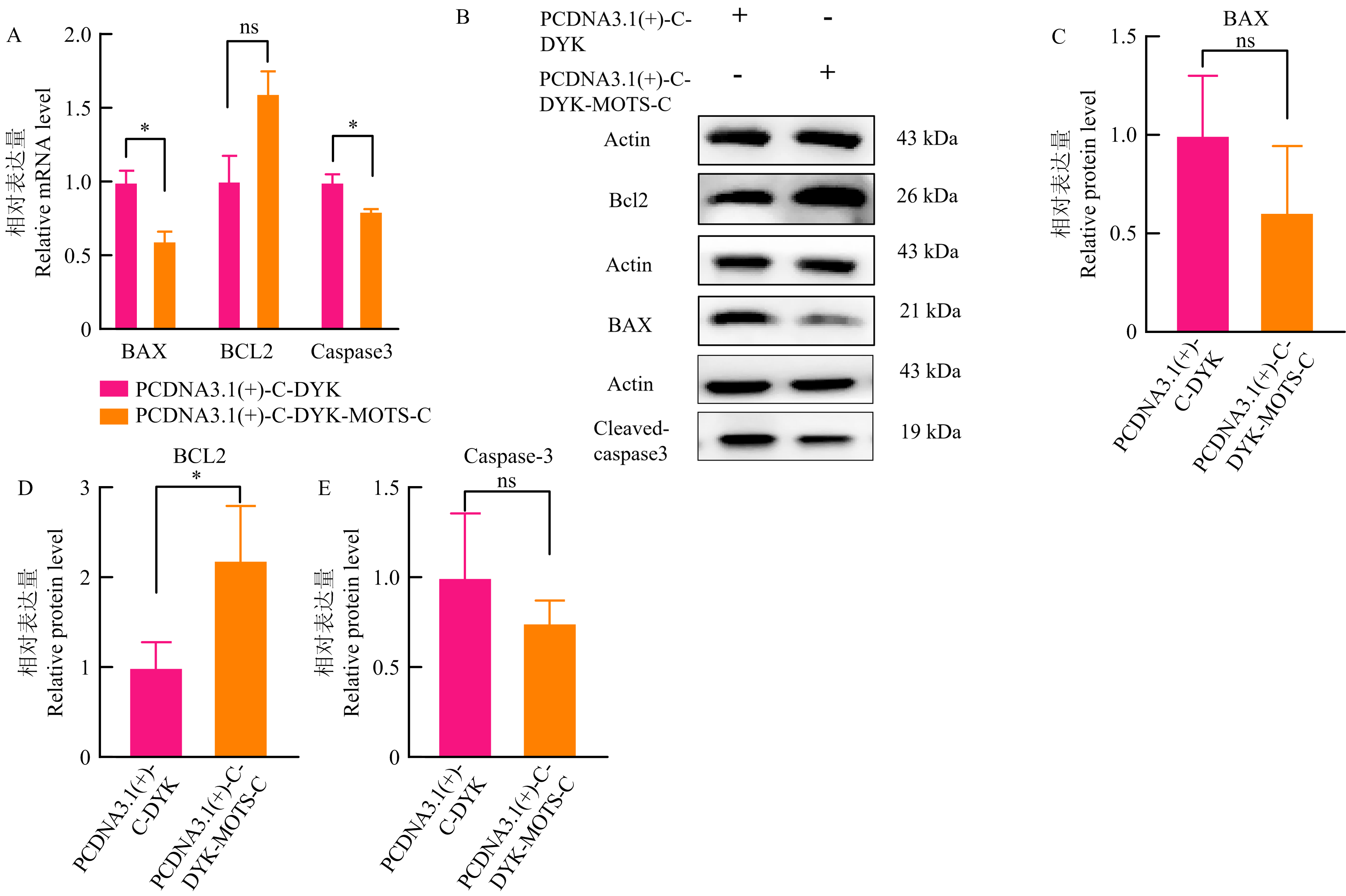
Effects of MOTS-C overexpression on apoptosis in IPEC-J2 cells A: Changes in mRNA expression levels of apoptosis-related genes after transfection with the MOTS-C overexpression plasmid for 24 h; B-E: Changes in protein expression levels of apoptosis-related proteins were analyzed 48 h after transfection with the MOTS-C overexpression plasmid for 48 h. Experiments were repeated three times, n = 3"

Fig. 7

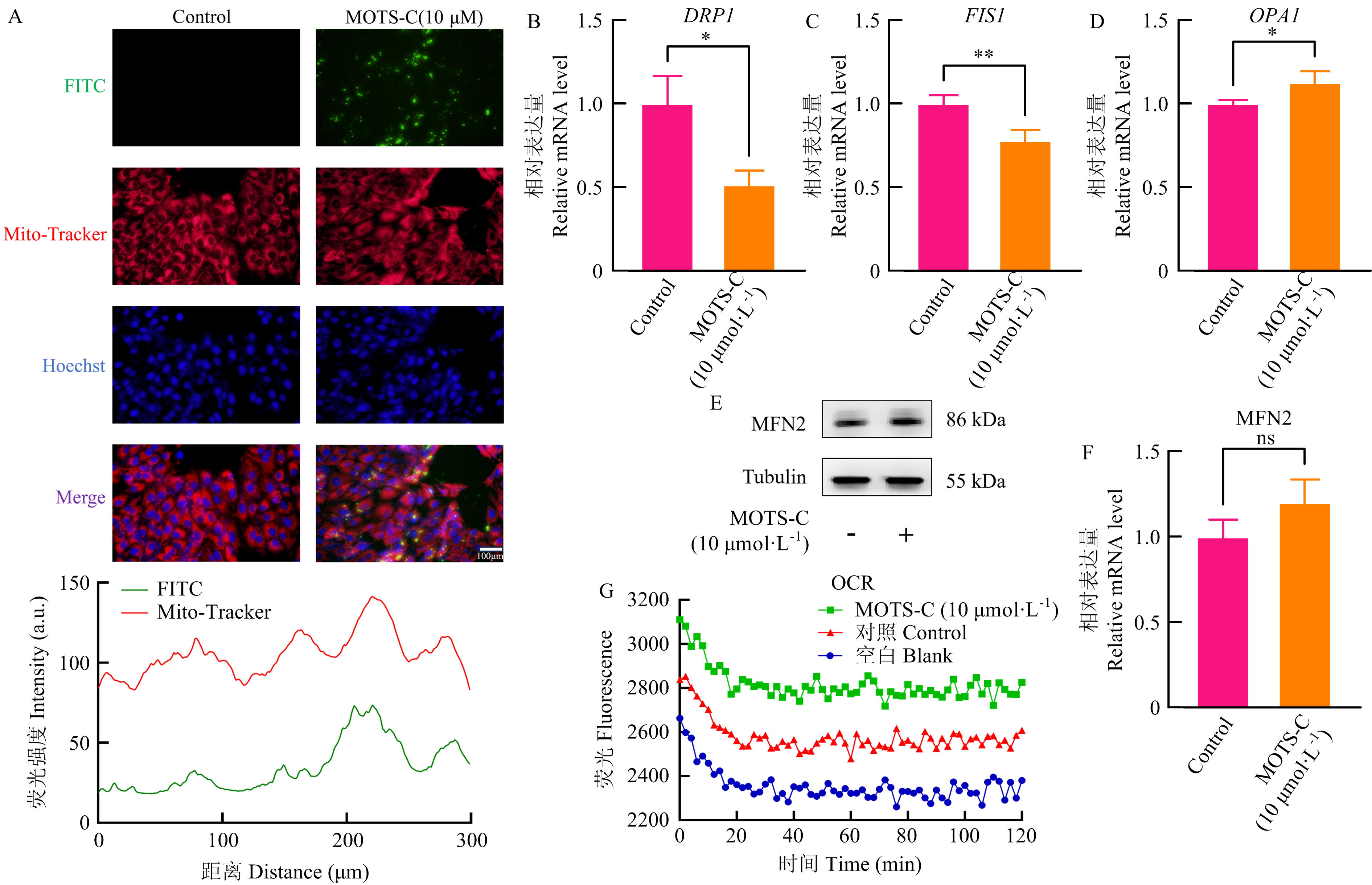
Co-localization of exogenous MOTS-C peptide with mitochondria and its effects on mitochondrial function in IPEC-J2 cells A: The co-localization MOTS-C with mitochondria after the exogenous MOTS-C peptide treatment for 24 h(A); B-F: Changes in mRNA levels (B-D) and protein levels (E-F) of mitochondrial-related genes after the exogenous MOTS-C peptide treatment for 24 h; G: OCR detection. Experiments were repeated three times, n = 3"

Fig. 8

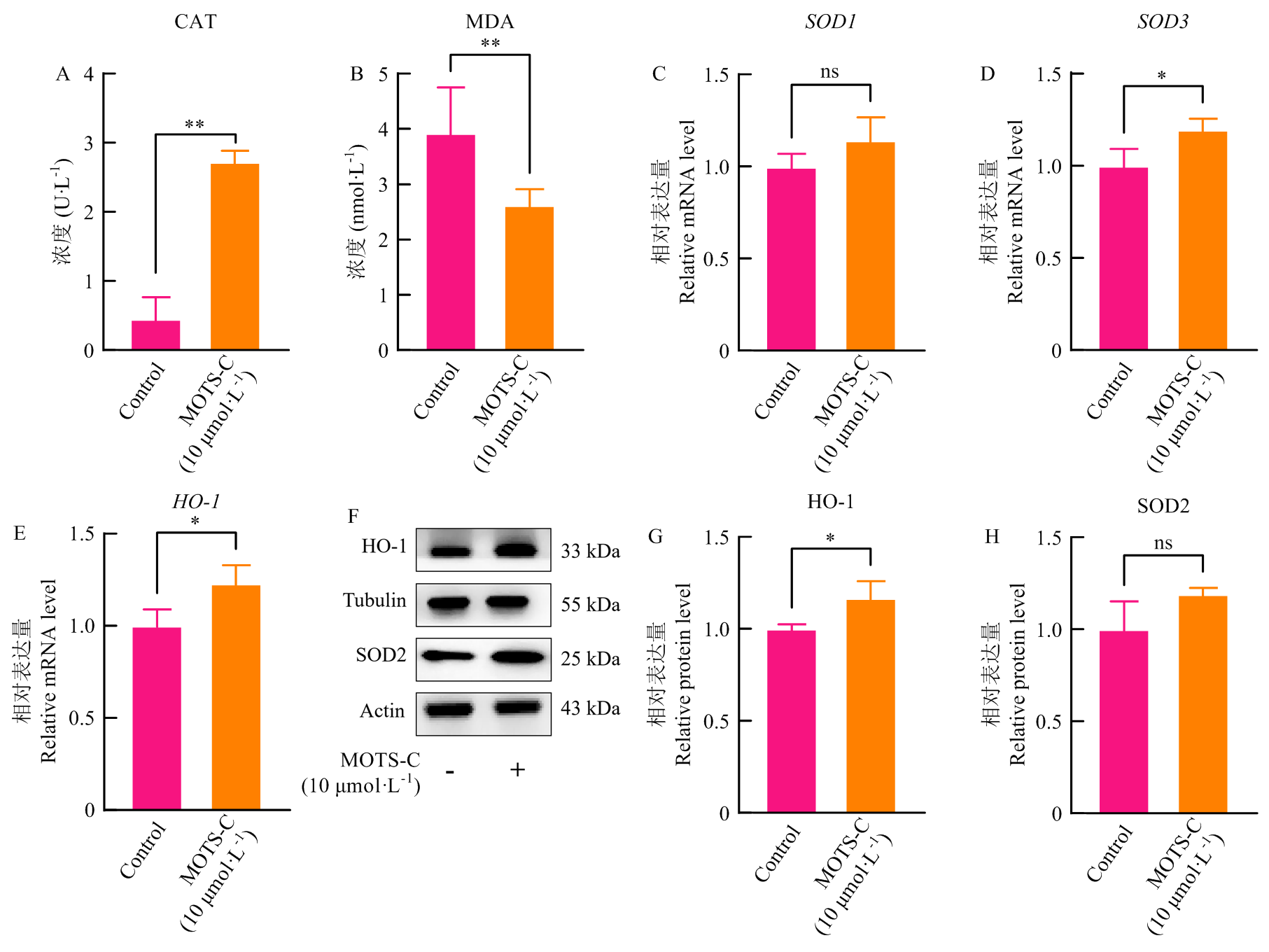
Effects of exogenous MOTS-C peptide on oxidative stress in IPEC-J2 cells Intracellular oxidative stress indicators were detected using assay kits (A-B), mRNA expression levels of intracellular antioxidant genes were detected using qRT-PCR (C-E) and Western blot (F-H) after the exogenous MOTS-C peptide treatment for 24 h. Experiments were repeated three times, n = 3"

Fig. 9

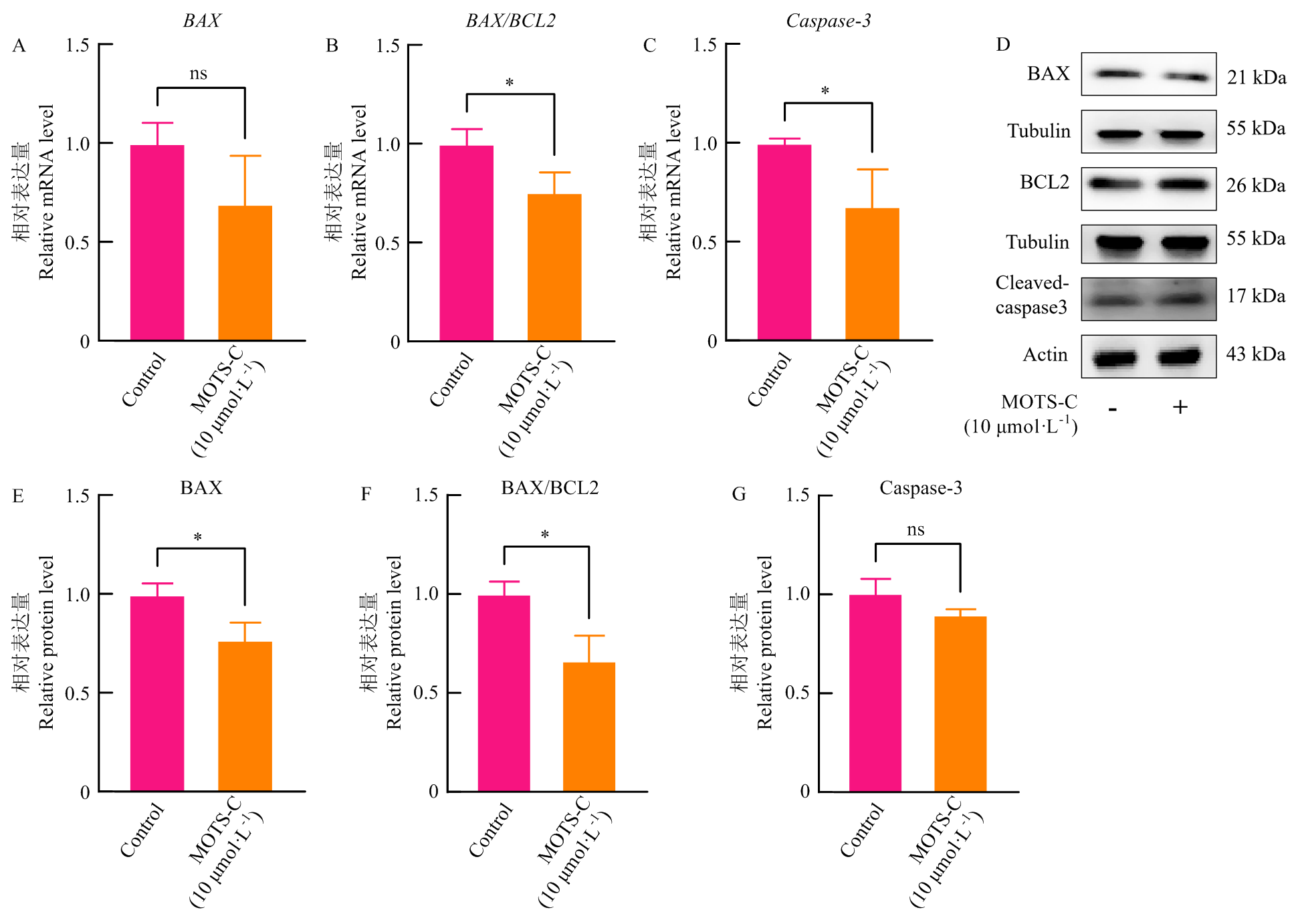
Effects of exogenous MOTS-C peptide on apoptosis in IPEC-J2 cells Changes of apoptosis-related genes in mRNA (A-C) and protein (D-G) levels after the exogenous MOTS-C peptide treatment for 24 h. Experiments were repeated three times, n = 3"

[1]
Li Y, Li Z Z, Ren Y Y, Lei Y, Yang S L, Shi Y Q, Peng H, Yang W J, Guo T T, Yu Y, Xiong Y Y. Mitochondrial-derived peptides in cardiovascular disease: Novel insights and therapeutic opportunities[J]. Journal of Advanced Research, 2024, 64: 99-115.

doi: 10.1016/j.jare.2023.11.018
[2]
Moschandrea C, Kondylis V, Evangelakos I, Herholz M, Schneider F, Schmidt C, Yang M, Ehret S, Heine M, Jaeckstein M Y, Szczepanowska K, Schwarzer R, Baumann L, Bock T, Nikitopoulou E, Brodesser S, Krüger M, Frezza C, Heeren J, Trifunovic A, et al. Mitochondrial dysfunction abrogates dietary lipid processing in enterocytes[J]. Nature, 2024, 625(7994): 385-392.

doi: 10.1038/s41586-023-06857-0
[3]
Lee C, Zeng J, Drew B G, Sallam T, Martin-Montalvo A, Wan J X, Kim S J, Mehta H, Hevener A L, de Cabo R, Cohen P. The mitochondrial-derived peptide MOTS-c promotes metabolic homeostasis and reduces obesity and insulin resistance[J]. Cell Metabolism, 2015, 21(3): 443-454.

doi: 10.1016/j.cmet.2015.02.009 pmid: 25738459
[4]
Zhang W L, Li S P, Zhang Y L, Wu Y X, Chen D, Pang Q F, Han S G. MOTS-c attenuates airway barrier dysfunction in allergic asthma by inhibiting epithelial apoptosis via Nrf2 pathway[J]. International Immunopharmacology, 2025, 161: 115014.

doi: 10.1016/j.intimp.2025.115014
[5]
Zarse K, Ristow M. A mitochondrially encoded hormone ameliorates obesity and insulin resistance[J]. Cell Metabolism, 2015, 21(3): 355-356.

doi: 10.1016/j.cmet.2015.02.013 pmid: 25738453
[6]
Yong C Q Y, Tang B L. A mitochondrial encoded messenger at the nucleus[J]. Cells, 2018, 7(8): 105.

doi: 10.3390/cells7080105
[7]
Reynolds J C, Lai R W, Woodhead J S T, Joly J H, Cameron-Smith D, Lu R, Cohen P, Graham N A, Merry T L, Lee C. MOTS-c is an exercise-induced mitochondrial-encoded regulator of age-dependent physical decline and muscle homeostasis[J]. Nature Communications, 2021, 12: 470.

doi: 10.1038/s41467-020-20790-0 pmid: 33473109
[8]
Yuan J H, Wang M D, Pan Y R, Liang M, Fu Y, Duan Y M, Tang M, Laher I, Li S C. The mitochondrial signaling peptide MOTS-c improves myocardial performance during exercise training in rats[J]. Scientific Reports, 2021, 11: 20077.

doi: 10.1038/s41598-021-99568-3 pmid: 34635713
[9]
Kim K H, Son J M, Benayoun B A, Lee C. The mitochondrial- encoded peptide MOTS-c translocates to the nucleus to regulate nuclear gene expression in response to metabolic stress[J]. Cell Metabolism, 2018, 28(3): 516-524.e7.

doi: 10.1016/j.cmet.2018.06.008
[10]
Tang M, Su Q S, Duan Y M, Fu Y, Liang M, Pan Y R, Yuan J H, Wang M D, Pang X L, Ma J C, Laher I, Li S C. The role of MOTS-c-mediated antioxidant defense in aerobic exercise alleviating diabetic myocardial injury[J]. Scientific Reports, 2023, 13: 19781.

doi: 10.1038/s41598-023-47073-0
[11]
Feng Y W, Rao Z J, Tian X, Hu Y, Yue L T, Meng Y F, Zhong Q L, Chen W, Xu W L, Li H R, Hu Y J, Shi R F. Endurance training enhances skeletal muscle mitochondrial respiration by promoting MOTS-c secretion[J]. Free Radical Biology and Medicine, 2025, 227: 619-628.

doi: 10.1016/j.freeradbiomed.2024.12.038 pmid: 39706498
[12]
Li F F, Jia Y, Fang J, Gong L Q, Zhang Y Z, Wei S S, Wu L L, Jiang P. Neuroprotective mechanism of MOTS-c in TBI mice: Insights from integrated transcriptomic and metabolomic analyses[J]. Drug Design, Development and Therapy, 2024, 18: 2971-2987.

doi: 10.2147/DDDT.S460265 pmid: 39050800
[13]
Yin X Q, Jing Y Y, Chen Q, Abbas A B, Hu J L, Xu H M. The intraperitoneal administration of MOTS-c produces antinociceptive and anti-inflammatory effects through the activation of AMPK pathway in the mouse formalin test[J]. European Journal of Pharmacology, 2020, 870: 172909.

doi: 10.1016/j.ejphar.2020.172909
[14]
Zhai D S, Ye Z C, Jiang Y H, Xu C M, Ruan B J, Yang Y, Lei X Y, Xiang A, Lu H Y, Zhu Z, Yan Z, Wei D, Li Q Y, Wang L, Lu Z F. MOTS-c peptide increases survival and decreases bacterial load in mice infected with MRSA[J]. Molecular Immunology, 2017, 92: 151-160.

doi: S0161-5890(17)30546-1 pmid: 29096170
[15]
Jiang J H, Chang X, Nie Y Y, Shen Y X, Liang X Y, Peng Y L, Chang M. Peripheral administration of a cell-penetrating MOTS-c analogue enhances memory and attenuates Aβ1-42- or LPS-induced memory impairment through inhibiting neuroinflammation[J]. ACS Chemical Neuroscience, 2021, 12(9): 1506-1518.

doi: 10.1021/acschemneuro.0c00782
[16]
Barszcz M, Skomiał J. The development of the small intestine of piglets - chosen aspects[J]. Journal of Animal and Feed Sciences, 2011, 20(1): 3-15.

doi: 10.22358/jafs/66152/2011
[17]
Sabeena Farvin K H, Andersen L L, Otte J, Nielsen H H, Jessen F, Jacobsen C. Antioxidant activity of cod (Gadus morhua) protein hydrolysates: Fractionation and characterisation of peptide fractions[J]. Food Chemistry, 2016, 204: 409-419.

doi: S0308-8146(16)30322-3 pmid: 26988519
[18]
Suetsuna K, Ukeda H, Ochi H. Isolation and characterization of free radical scavenging activities peptides derived from casein[J]. The Journal of Nutritional Biochemistry, 2000, 11(3): 128-131.

doi: 10.1016/S0955-2863(99)00083-2
[19]
Keskin O, Gursoy A, Ma B Y, Nussinov R. Principles of Protein- Protein interactions: What are the preferred ways for proteins to interact?[J]. Chemical Reviews, 2008, 108(4)1225-1244.

doi: 10.1021/cr040409x pmid: 18355092
[20]
van der Bliek A M, Shen Q, Kawajiri S. Mechanisms of mitochondrial fission and fusion[J]. Cold Spring Harbor Perspectives in Biology, 2013, 5(6): a011072.
[21]
Adebayo M, Singh S, Singh A P, Dasgupta S. Mitochondrial fusion and fission: The fine-tune balance for cellular homeostasis[J]. FASEB Journal, 2021, 35(6): e21620.

doi: 10.1096/fsb2.v35.6
[22]
Song Z Y, Ghochani M, McCaffery J M, Frey T G, Chan D C. Mitofusins and OPA1 mediate sequential steps in mitochondrial membrane fusion[J]. Molecular Biology of the Cell, 2009, 20(15): 3525-3532.

doi: 10.1091/mbc.E09-03-0252 pmid: 19477917
[23]
von der Malsburg A, Sapp G M, Zuccaro K E, von Appen A, Moss F R, Kalia R, Bennett J A, Abriata L A, Dal Peraro M, van der Laan M, Frost A, Aydin H. Structural mechanism of mitochondrial membrane remodelling by human OPA1[J]. Nature, 2023, 620(7976): 1101-1108.

doi: 10.1038/s41586-023-06441-6
[24]
Chen T, Shuang R N, Gao T T, Ai L J, Diao J C, Yuan X Y, He L, Tao W W, Huang X. OPA1 mediated fatty acid β-oxidation in hepatocyte: The novel insight for melatonin attenuated apoptosis in concanavalin a induced acute liver injury[J]. Journal of Pineal Research, 2024, 76(8): e70010.

doi: 10.1111/jpi.v76.8
[25]
Noone J, O’Gorman D J, Kenny H C. OPA1 regulation of mitochondrial dynamics in skeletal and cardiac muscle[J]. Trends in Endocrinology & Metabolism, 2022, 33(10): 710-721.
[26]
Roe A J, Qi X. Drp1 phosphorylation by MAPK1 causes mitochondrial dysfunction in cell culture model of Huntington’s disease[J]. Biochemical and Biophysical Research Communications, 2018, 496(2): 706-711.

doi: 10.1016/j.bbrc.2018.01.114
[27]
Kleele T, Rey T, Winter J, Zaganelli S, Mahecic D, Perreten Lambert H, Ruberto F P, Nemir M, Wai T, Pedrazzini T, Manley S. Distinct fission signatures predict mitochondrial degradation or biogenesis[J]. Nature, 2021, 593(7859): 435-439.

doi: 10.1038/s41586-021-03510-6
[28]
Sreekumar P G, Ishikawa K, Spee C, Mehta H H, Wan J X, Yen K, Cohen P, Kannan R, Hinton D R. The mitochondrial-derived peptide humanin protects RPE cells from oxidative stress, senescence, and mitochondrial dysfunction[J]. Investigative Ophthalmology & Visual Science, 2016, 57(3): 1238-1253.
[29]
Matsunaga D, Sreekumar P G, Ishikawa K, Terasaki H, Barron E, Cohen P, Kannan R, Hinton D R. Humanin protects RPE cells from endoplasmic reticulum stress-induced apoptosis by upregulation of mitochondrial glutathione[J]. PLoS ONE, 2016, 11(10): e0165150.

doi: 10.1371/journal.pone.0165150
[30]
Kannan H T, Umapathy S, Pan I. SHLP6: A novel NLRP3 and Cav1 modulating agent in Cu-induced oxidative stress and neurodegeneration[J]. Frontiers in Molecular Neuroscience, 2025, 18: 1553308.

doi: 10.3389/fnmol.2025.1553308
[31]
Li X Y, Zhan F L, Qiu G F, Lu P, Shen Z H, Qi Y P, Wu M C, Chu M Y, Feng J, Wen Z A, Yao X, Wang A, Jin W J, Zhang X, Liao J J, Zhang J L, Song M J, Wang W, Wang X W. MOTS-c attenuates lung ischemia-reperfusion injury via MYH9-Dependent nuclear translocation and transcriptional activation of antioxidant genes[J]. Redox Biology, 2025, 84: 103681.

doi: 10.1016/j.redox.2025.103681
[32]
Morris D L, Kastner D W, Johnson S, Strub M P, He Y, Bleck C K E, Lee D Y, Tjandra N. Humanin induces conformational changes in the apoptosis regulator BAX and sequesters it into fibers, preventing mitochondrial outer-membrane permeabilization[J]. Journal of Biological Chemistry, 2019, 294(50): 19055-19065.

doi: 10.1074/jbc.RA119.011297 pmid: 31690630
[33]
Morris D L, Johnson S, Bleck C K E, Lee D Y, Tjandra N. Humanin selectively prevents the activation of pro-apoptotic protein BID by sequestering it into fibers[J]. The Journal of Biological Chemistry, 2020, 295(52): 18226-18238.

doi: 10.1074/jbc.RA120.013023
[34]
Cobb L J, Lee C, Xiao J L, Yen K, Wong R G, Nakamura H K, Mehta H H, Gao Q L, Ashur C, Huffman D M, Wan J X, Muzumdar R, Barzilai N, Cohen P. Naturally occurring mitochondrial-derived peptides are age-dependent regulators of apoptosis, insulin sensitivity, and inflammatory markers[J]. Aging, 2016, 8(4): 796-809.

doi: 10.18632/aging.v8i4
[35]
Yin Y D, Li Y J, Ma B Y, Ren C L, Zhao S H, Li J, Gong Y, Yang H, Li J B. Mitochondrial-derived peptide MOTS-c suppresses ovarian cancer progression by attenuating USP7-mediated LARS1 deubiquitination[J]. Advanced Science, 2024, 11(43): e2405620.
[36]
Zhang Y L, Huang J F, Zhang Y R, Jiang F J, Li S P, He S, Sun J J, Chen D, Tong Y, Pang Q F, Wu Y X. The Mitochondrial-Derived Peptide MOTS-c Alleviates Radiation Pneumonitis via an Nrf2- Dependent Mechanism[J/OL]. Antioxidants, 2024, 13(5): 613. https://doi.org/10.3390/antiox13050613.
[37]
Zhong P, Peng J Y, Hu Y W, Zhang J, Shen C J. Mitochondrial derived peptide MOTS-c prevents the development of heart failure under pressure overload conditions in mice[J]. Journal of Cellular and Molecular Medicine, 2022, 26(21): 5369-5378.

doi: 10.1111/jcmm.v26.21
[1] GERIQIMUGE, PUBUZHANDUI, XU Qing, HOU LingLing. Effects of Hypoxia on Proliferation of Bovine Renal Cells and Mitochondrial Autophagy [J]. Scientia Agricultura Sinica, 2026, 59(6): 1333-1347.
[2] MA GuiLan, ZHANG XuYang, LI Wu. Regulatory Role of Guanylate-Binding Protein 2 in Staphylococcus aureus -Induced Macrophage Apoptosis [J]. Scientia Agricultura Sinica, 2026, 59(4): 912-926.
[3] SUN ZhenYu, CUI YiGe, LI YuQi, WANG SiQi, DU Xing, LI QiFa. Transcription Factor ZBTB6 Activates the Transcription and Function of NORHA in Sow Granulosa Cells [J]. Scientia Agricultura Sinica, 2026, 59(13): 2962-2974.
[4] WANG Wei, LUO ChunHai, JIA HongDou, LIU JiaJin, LI DanYang, FU ShiXin. Effect of Gln on Endoplasmic Reticulum Stress in Retained Fetal Membranes Cows Under Oxidative Stress via the PI3K/AKT Pathway [J]. Scientia Agricultura Sinica, 2025, 58(7): 1451-1462.
[5] LI RuiTong, CHEN QingMei, XU JianFang, ZHANG JunMin, SI Wei, ZHANG TieYing. Study on the Antioxidant Function and Mechanism of 18β-glycyrrhetinic Acid Using Porcine Intestinal Organoid Model [J]. Scientia Agricultura Sinica, 2025, 58(22): 4771-4785.
[6] LI XiangYu, LIU JianZhuo, HU DanDan, LIU GengYu, CHEN LiangYu, LI Bing, DU WanLi, SONG Bo. Characterization of Maize Germplasm Resistance to Common Smut and Analysis of Physiological Differences [J]. Scientia Agricultura Sinica, 2025, 58(13): 2504-2521.
[7] WANG SiQi, ZHOU ChunXue, LI YuQi, DU Xing, PAN ZengXiang, LI QiFa. miR-34a Induces Early Apoptosis of Porcine Ovarian Granulosa Cells by Activating lncRNA NORHA Transcription [J]. Scientia Agricultura Sinica, 2024, 57(5): 1000-1009.
[8] MENG ZhaoYi, WANG YunLu, YAO YiLong, XI GuangYin, NIU JiaQiang, SOLANGSIZHU, GUO Min, XU YeFen. lncRNA-MSTRG.7889.1 Competitively Binds to bta-miR-146a Targeting Smad4 to Regulate Apoptosis of Yak Granulosa Cells [J]. Scientia Agricultura Sinica, 2024, 57(4): 797-809.
[9] ZHANG Bing, YANG YanYan, FENG QianHui, SHI Wen, FANG YiZhen, HUANG JiaBao, SHI DeShun. Effects of Sodium Selenite on the in vitro Maturation of Porcine Oocytes and Their Embryonic Development Potentials [J]. Scientia Agricultura Sinica, 2024, 57(17): 3482-3493.
[10] WANG JinPeng, LUORENG ZhuoMa, LI YanXia, FENG Fen, WANG ZhengXing, PAN ChuanYing, LAN XianYong, WANG XingPing. The Function of lncRNA RRAS2-AS1 in LPS Induced Bovine Mammary Epithelial Cells Inflammation [J]. Scientia Agricultura Sinica, 2024, 57(14): 2874-2888.
[11] PAN YangYang, WANG JingLei, WANG Meng, WANG LiBin, ZHANG Qian, CHEN Rui, ZHANG TianTian, CUI Yan, XU GengQuan, FAN JiangFeng, YU SiJiu. Formation and Function of Paraspeckle During Pre-implantation Embryos Development in Yak [J]. Scientia Agricultura Sinica, 2023, 56(6): 1189-1203.
[12] CUI HongJie, LU ChunTing, PAN LiQin, HU Hui, ZHONG PeiYun, ZHU JieYing, ZHANG KaiZhao, HUANG XiaoHong. Curcumin Alleviates Zearalenone-Induced Oxidative Damage in Porcine Renal Epithelial Cells via SIRT1/FOXO1 Pathway [J]. Scientia Agricultura Sinica, 2023, 56(5): 1007-1018.
[13] XI MengXue, SHEN Dan, SHI YiFan, LI ChunMei. Effects of TBHQ on Pyroptosis, Necroptosis and Inflammatory Damage of Chicken Embryonic Lung Tissues Induced by PM2.5 from Chicken Houses [J]. Scientia Agricultura Sinica, 2023, 56(4): 779-787.
[14] TAO WenJing, ZHANG ZiTing, LIU Yuan, SONG Dan, LI XiangChen. Inhibitory Effect of N-acetylcysteine on Bisphenol A-Induced Apoptosis and Inflammatory Response in Porcine Kidney Cells [J]. Scientia Agricultura Sinica, 2023, 56(3): 549-558.
[15] ZHANG Nan, HAN GuangJie, LIU Qin, LI ChuanMing, QI JianHang, LU YuRong, XIA Yang, XU Jian. Response and Function of the Transcription Factor CncC in Cnaphalocrocis medinalis Infected with Baculovirus CnmeGV [J]. Scientia Agricultura Sinica, 2023, 56(13): 2491-2503.
Viewed
Full text


Abstract

Cited

  Shared   
  Discussed   
No Suggested Reading articles found!