Scientia Agricultura Sinica ›› 2025, Vol. 58 ›› Issue (4): 819-830.doi: 10.3864/j.issn.0578-1752.2025.04.015

• RESEARCH NOTES • Previous Articles    

Effect of FGF5 and FGF21 on Proliferation of Dermal Papilla Cells in Cashmere Goat

WANG Niu(), SHI XinRan, ZHANG WeiDong, WANG Xin()   

  1. College of Animal Science and Technology, Northwest A&F University, Yangling 712100, Shaanxi
  • Received:2023-08-01 Accepted:2024-12-30 Online:2025-02-16 Published:2025-02-24
  • Contact: WANG Xin

Abstract:

【Background】The hair follicle cycle in goat is divided into three phases: the growth phase (anagen), the regression phase (catagen), and the resting phase (telogen). Fibroblast growth factor 5 (FGF5) and fibroblast growth factor 21 (FGF21) are important regulators in goat hair cycle. However, the precise localization of FGF5 and FGF21 in goat skin tissue and their regulatory mechanisms remain unclear during hair follicle cycle transition. Dermal papilla cells (DPCs) located in the dermal papilla region, are essential for hair follicle development. When DPCs lose their function, hair follicle remains in the telogen, leading to hair loss.【Objective】Therefore, this study aimed to identify the expression and localization of FGF5 and FGF21 in Cashmere goat skin tissue, understand their roles in DPCs, and to analyze their regulatory mechanisms. This would enrich the knowledge of fibroblast growth factor family proteins in hair follicle cycle regulation and provide a theoretical basis for further elucidating the molecular mechanisms of Cashmere goat hair follicle cycle transition. 【Method】Real-time quantitative PCR (RT-qPCR) and enzyme-linked immunosorbent assay (ELISA) were used to measure the expression levels of FGF5, FGF21 and their receptor 1 (FGFR1) in the skin tissue of Cashmere goats at anagen and telogen. Single-cell transcriptome sequencing data were analyzed to determine the expression and localization of FGF5, FGF21 and FGFR1, which were validated through immunofluorescence methods. Adenovirus was explored to overexpress FGF5 and FGF21 in DPCs. The effects on DPCs proliferation were measured using MTT, EdU and flow cytometry. The expressions of FGFR1 and proliferation-related genes were detected using RT-qPCR, immunofluorescence and western blotting methods. RT-qPCR, immunofluorescence and western blotting were used to investigate the effect of FGF5 and FGF21 on the proliferation of DPCs. 【Result】FGF5, FGF21 and FGFR1 exhibited higher expression levels in anagen than that in telogen, all of which were located within DPCs. Both FGF5 and FGF21 restrained DPCs viability, which significantly reduced the number of EdU-positive cells. FGF5 decreased the percentage of DPCs in G2 and S phases, while FGF21 specifically affected the G2 phase. The expression of FGFR1 was significantly increased following the overexpression of FGF5 and FGF21. Moreover, the expressions of cell proliferation markers KI67 and PCNA were also significantly inhibited with the overexpression of FGF5 and FGF21. Additionally, the expression of β-catenin, a key component of Wnt/β-catenin signaling pathway, was reduced. Thereby the activity of transcription factors TCF3 and JUN was inhibited, which were the downstream effectors of Wnt/β-catenin signaling pathway. Thus the inhibition further led to the decreased expression of proliferation-related genes MYC and CYCLIND1. 【Conclusion】FGF5 and FGF21 played a critical role as regulatory factors in controlling the biological function of DPCs. This study provided the first evidences that FGF21, similar to FGF5, restrained the proliferation of DPCs by suppressing the Wnt/β-catenin signaling pathway. These findings contributes to our understanding of how fibroblast growth factors influence hair follicle cycle transition, particularly in the context of Cashmere goat hair growth and cashmere production.

Key words: cashmere goat, FGF5, FGF21, DPCs, proliferation, Wnt/β-catenin signaling pathway

Table 1

Information of primers for RT-qPCR"

基因名称
Gene symbol
GenBank登录号
Accession No.
引物序列
Primer sequence (5′-3′)
产物长度
Production size (bp)
退火温度
Tm (℃)
β-actin NM_001314342.1 5′-GCAGACAGGATGCAGAAAGA-3′ 96 56
5′-GCAAGTACTCCGTGTGGATT-3′
FGF5 NM_001291973.1 5′-CCGCGATACACAGAACTGAA-3′ 129 58
5′-ATCTTGGCAGAAAGTGGGTAG-3′
FGF21 XM_005692688.3 5′-GCGGGTCTGTCTGGATATAAA-3′ 118 62
5′-CTCAGATGGCTGCTTCAGTA -3′
FGFR1 XM_018041774.1 5′-GAGGCTACAAGGTCCGTTATG-3′ 125 62
5′-GAGCTGGTAGGTATGGTTGATG-3′
FGFR3 XM_018045593.1 5′-GTCCGAGATGGAGATGATGAAG-3′ 103 58
5′-GTACTCCACCAGCACATACAG-3′
CTNNB1 XM_018066894.1 5′-CTGGACTCTGGAATCCATTCTG-3′ 103 59
5′-GATACCACCCAAGTCCTGTATG-3′
C-JUN XM_018044742.1 5′-CCAAGAACTGCATGGACCTAA-3′ 111 56
5′-AGATCGCTTCTGTAGTACTCCT-3′
C-MYC XM_018058563.1 5′-GAAGAGGCGAGAACAGTTGAA-3′ 86 58
5′-CTATTGGAGGGAGGAACTGAAC-3′
CCND1 XM_018043271.1 5′-GCAGTCTTAGGCATCCTGT-3′ 131 58
5′-CCTAGCCGAGAGGTTACAT-3′
PCNA XM_005688167.3 5′-GCAGACGCGGCAGTATTA-3′ 107 62
5′-CTCATCCTCTGAAGCCTAAGC-3′
MKI67 XM_005698601.3 5′-CTCAGGAAGCCTACACATACAC-3′ 108 58
5′-AGCATCTTGGGAAACTCCAG-3′
TCF3 XM_018050411.1 5′-CCTGACTCCTATGGTGATCTTG-3′ 131 58
5′-AAGAAGGACCTCAAAGCTCC-3′

Table 2

Antibody information"

抗体名称 Name of antibody 生产厂商 Manufacturer 货号 Cat No.
FGF5 Polyclonal Antibody Proteintech 18171-1-AP
FGF21 Polyclonal Antibody Proteintech 26272-1-AP
FGFR1 Monoclonal Antibody Proteintech 60325-1-Ig
GAPDH Polyclonal Antibody Proteintech 10494-1-AP
Beta Catenin Polyclonal Antibody Proteintech 51067-2-AP
C-MYC Polyclonal Antibody Proteintech 10828-1-AP
Cyclin D1 Polyclonal Antibody Proteintech 26939-1-AP
PCNA Polyclonal Antibody Proteintech 24036-1-AP
Goat Anti-Rabbit IgG H&L (Alexa Fluor® 555) Abcam ab150078
Goat Anti-Mouse IgG H&L (Alexa Fluor® 647) Abcam ab150115

Fig. 1

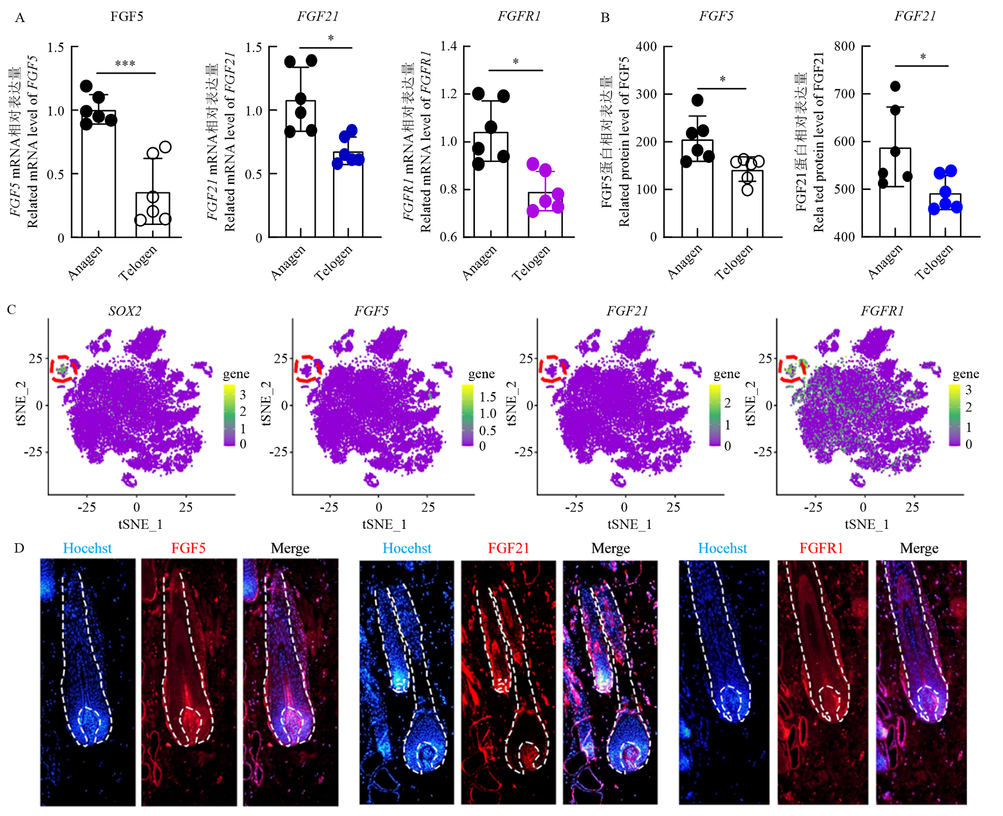
The expression pattern of FGF5 and FGF21 in cashmere goat dorsal skin A: Related mRNA level of FGF5, FGF21, and FGFR1 were detected by RT-qPCR (n=3); *P<0.05, ***P<0.001. B: Protein level of FGF5 and FGF21 were detected by ELISA (n=3); *P<0.05. C: Visualization of FGF5, FGF21, and FGFR1 expression across all single cells in tSNE plot. SOX2 were used to mark dermal papillae population. D: Immunostaining of FGF5, FGF21, and FGFR1 in hair follicle"

Fig. 2

The overexpression efficiency of FGF5 and FGF21 A: Related protein level of FGF5 and FGF21 was detected by ELISA (n=1). DPCs-pAd indicated the relative protein level of FGF5 or FGF21 from DPCs after adenovirus stimulating. CS-pAd indicated the relative protein level of FGF5 or FGF21 from cell culture medium (CS) after adenovirus stimulating. B: Related mRNA level of FGF5 and FGF21 was detected by RT-qPCR (n=3); ***P<0.01. C: FGF5 and FGF21 were detected by immunofluorescence staining. eGFP indicated the infection efficiency of adenovirus. Scale bar: 50 µm"

Fig. 3

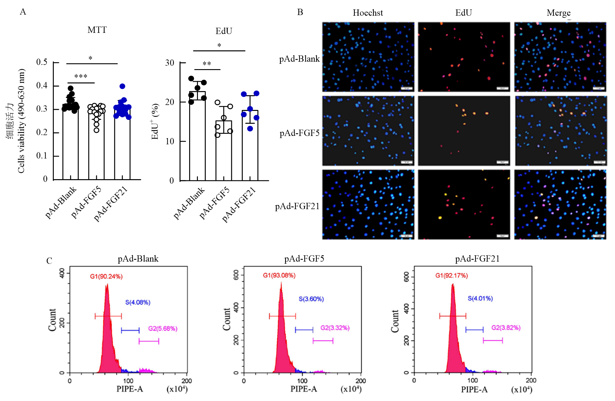
FGF5 and FGF21 inhibited the proliferation and cell cycle of DPCs A: Cell viability analysis of pAd-Black, pAd-FGF5, and pAd-FGF21 on DPCs proliferation (n=16); *P<0.05, ***P<0.001; B: EdU incorporation assay of DPCs (n=3), ** P<0.01, *** P<0.001;. C: Cell cycle analysis of DPCs after pAd-Black, pAd-FGF5 and pAd-FGF21 exposure (n=3)"

Fig. 4

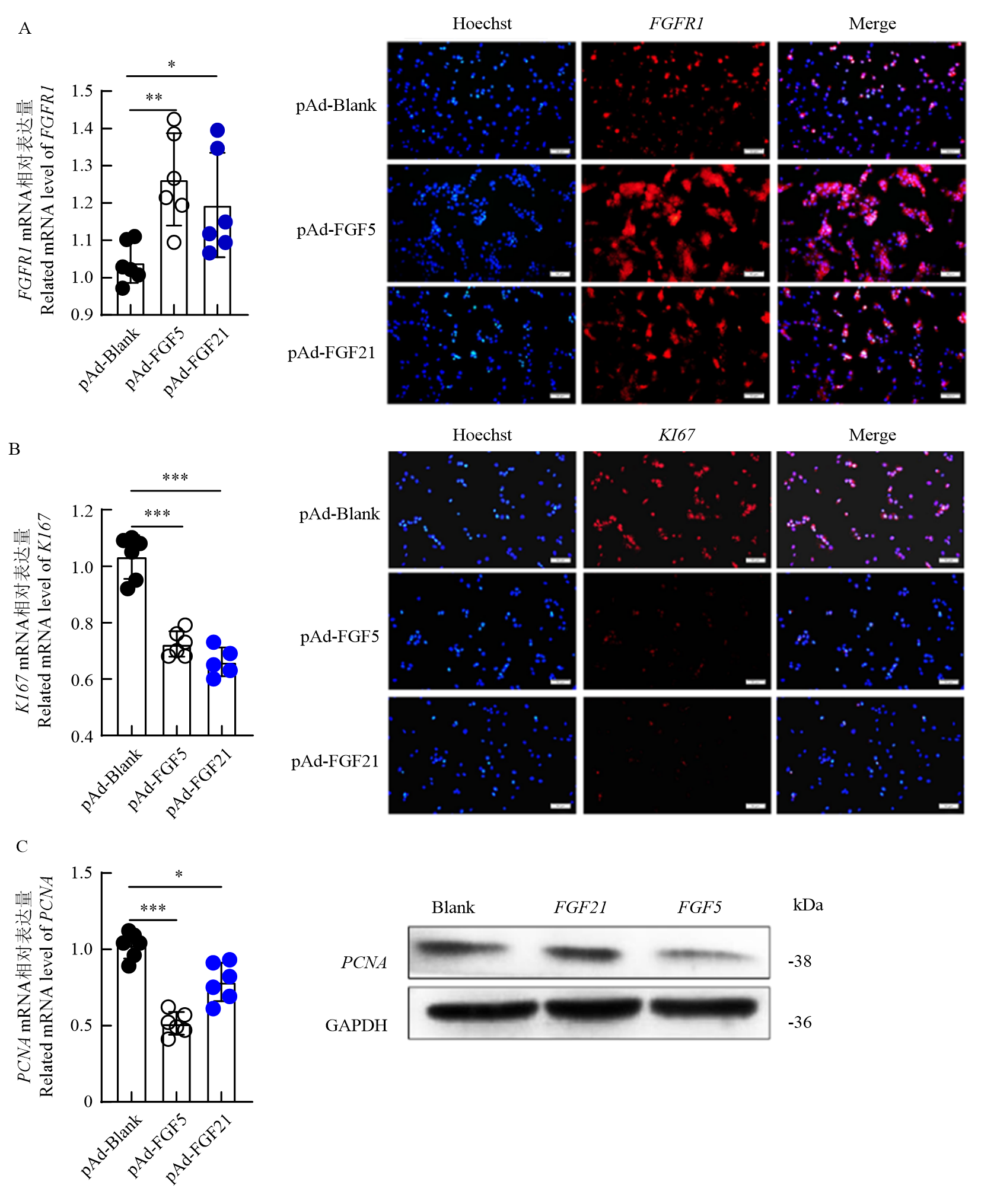
Effects of FGF5 and FGF21 on the expression of their receptor and proliferation-related genes A: The expression of FGFR1 was detected by RT-qPCR and immunofluorescence (n=3); *P<0.05, ** P<0.01; B: The expression of KI67 was detected by RT-qPCR and immunofluorescence (n=3); *** P<0.001; C: The expression of PCNA was detected by RT-qPCR and Western blotting (n=3); * P<0.05, *** P<0.001. Scale bar, 50 µm"

Fig. 5

FGF5 and FGF21 inhibited the expression of genes to Wnt/β-catenin signaling pathway A: Relative mRNA level of β-catenin, TCF3, JUN, MYC, and CYCLIND1 were detected by RT-qPCR (n=3); *P<0.05; ** P<0.01; *** P<0.001; B: Immunofluorescence staining results of β-catenin, MYC, and CYCLIND1. Scale bar: 50 µm; C: Western blotting results of β-catenin, MYC, and CYCLIND1"

Fig. 6

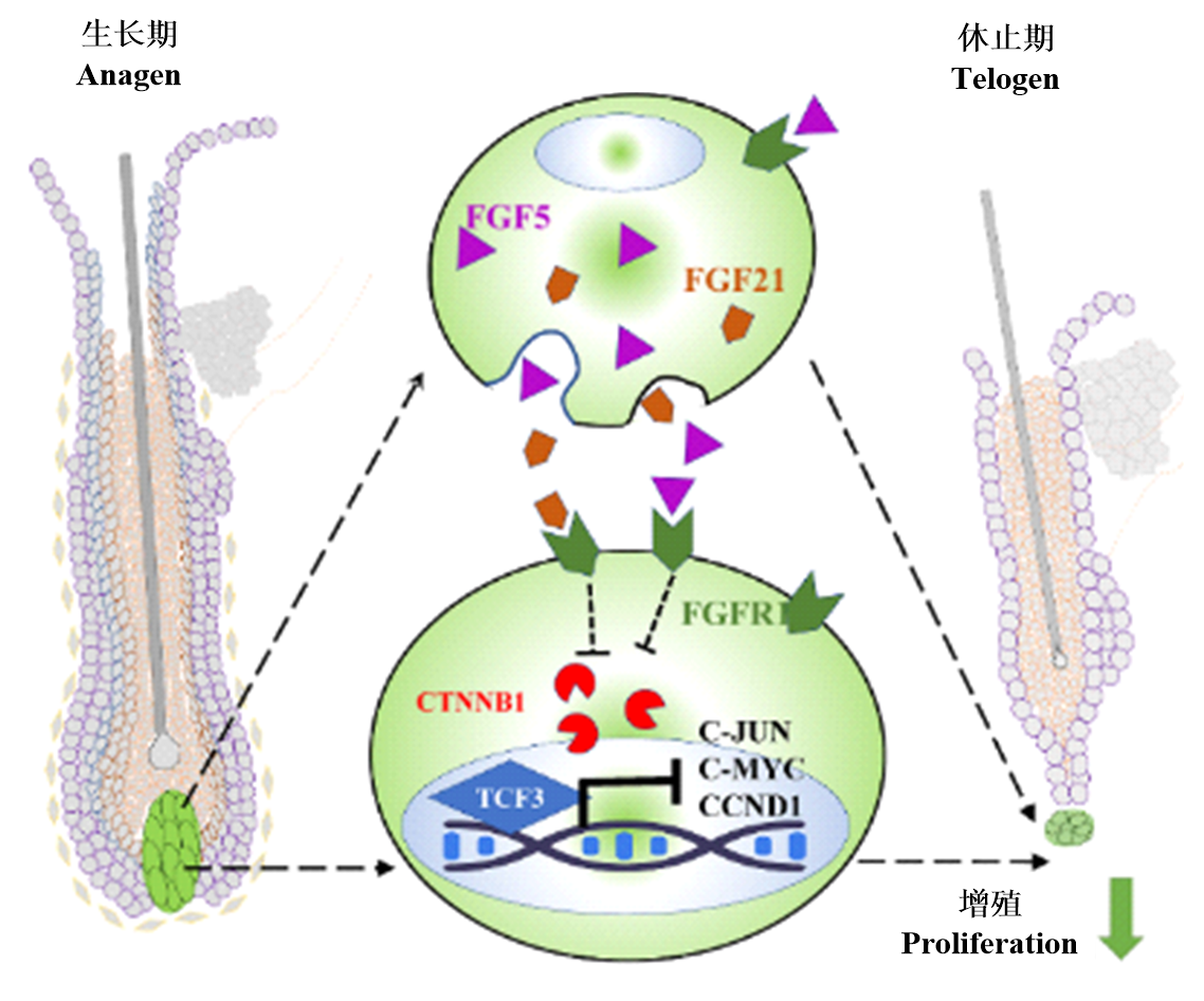
Effects of FGF5 and FGF21 on proliferation of DPCs in cashmere goat"

[1]
冯云奎, 王健, 马金亮, 张柳明, 李拥军. miR-31-5p对山羊毛囊干细胞增殖和凋亡的影响. 中国农业科学, 2021, 54(23): 5132-5143. doi: 10.3864/j.issn.0578-1752.2021.23.017.
FENG Y K, WANG J, MA J L, ZHANG L M, LI Y J. Effects of miR-31-5p on the proliferation and apoptosis of hair follicle stem cells in goat. Scientia Agricultura Sinica, 2021, 54(23): 5132-5143. doi: 10.3864/j.issn.0578-1752.2021.23.017. (in Chinese)
[2]
AVIGAD LARON E, AAMAR E, ENSHELL-SEIJFFERS D. The mesenchymal niche of the hair follicle induces regeneration by releasing primed progenitors from inhibitory effects of quiescent stem cells. Cell Reports, 2018, 24(4): 909-921.e3.

doi: S2211-1247(18)31011-8 pmid: 30044987
[3]
HE X L, CHAO Y, ZHOU G X, CHEN Y L. Fibroblast growth factor 5-short (FGF5s) inhibits the activity of FGF5 in primary and secondary hair follicle dermal papilla cells of Cashmere goats. Gene, 2016, 575(2): 393-398.
[4]
WANG X L, CAI B, ZHOU J K, ZHU H J, NIU Y Y, MA B H, YU H H, LEI A M, YAN H L, SHEN Q Y, SHI L, ZHAO X E, HUA J L, HUANG X X, QU L, CHEN Y L. Correction: disruption of FGF5 in Cashmere goats using CRISPR/Cas9 results in more secondary hair follicles and longer fibers. PLoS One, 2016, 11(11): e0167322.
[5]
MORGAN B A. The dermal papilla: an instructive niche for epithelial stem and progenitor cells in development and regeneration of the hair follicle. Cold Spring Harbor Perspectives in Medicine, 2014, 4(7): a015180.
[6]
ROMPOLAS P, DESCHENE E R, ZITO G, GONZALEZ D G, SAOTOME I, HABERMAN A M, GRECO V. Live imaging of stem cell and progeny behaviour in physiological hair-follicle regeneration. Nature, 2012, 487: 496-499.
[7]
CHI W, WU E, MORGAN B A. Dermal papilla cell number specifies hair size, shape and cycling and its reduction causes follicular decline. Development, 2013, 140(8): 1676-1683.

doi: 10.1242/dev.090662 pmid: 23487317
[8]
HWANG S B, PARK H J, LEE B H. Hair-growth-promoting effects of the fish collagen peptide in human dermal papilla cells and C57BL/6 mice modulating Wnt/β-catenin and BMP signaling pathways. International Journal of Molecular Sciences, 2022, 23(19): 11904.
[9]
HIGGINS C A, CHEN J C, CERISE J E, JAHODA C A B, CHRISTIANO A M. Microenvironmental reprogramming by three- dimensional culture enables dermal papilla cells to induce de novo human hair-follicle growth. Proceedings of the National Academy of Sciences of the United States of America, 2013, 110(49): 19679-19688.
[10]
GENTILE P, GARCOVICH S. Advances in regenerative stem cell therapy in androgenic alopecia and hair loss: Wnt pathway, growth- factor, and mesenchymal stem cell signaling impact analysis on cell growth and hair follicle development. Cells, 2019, 8(5): 466.
[11]
GRIGGS J, TRÜEB R M, GAVAZZONI DIAS M F R, HORDINSKY M, TOSTI A. Fibrosing alopecia in a pattern distribution. Journal of the American Academy of Dermatology, 2021, 85(6): 1557-1564.

doi: 10.1016/j.jaad.2019.12.056 pmid: 31926219
[12]
LIU X, ZHANG P, ZHANG X F, LI X, BAI Y, AO Y, HEXIG B, GUO X D, LIU D J. Fgf21 knockout mice generated using CRISPR/ Cas9 reveal genetic alterations that may affect hair growth. Gene, 2020, 733: 144242.
[13]
DONG Y, XIE M, JIANG Y, XIAO N Q, DU X Y, ZHANG W G, TOSSER-KLOPP G, WANG J H, YANG S, LIANG J, et al. Sequencing and automated whole-genome optical mapping of the genome of a domestic goat (Capra hircus). Nature Biotechnology, 2013, 31(2): 135-141.

doi: 10.1038/nbt.2478 pmid: 23263233
[14]
KAWANO M, KOMI-KURAMOCHI A, ASADA M, SUZUKI M, OKI J, JIANG J, IMAMURA T. Comprehensive analysis of FGF and FGFR expression in skin: FGF18 is highly expressed in hair follicles and capable of inducing anagen from telogen stage hair follicles. Journal of Investigative Dermatology, 2005, 124(5): 877-885.

pmid: 15854025
[15]
吴晋强, 曹校瑞, 闫瑞琴, 陆娜, 张娇娇, 吴佳豪, 赫晓燕. FGF21及其受体FGFR1和FGFR2在小鼠毛囊第1生长周期的表达. 畜牧兽医学报, 2019, 50(3): 534-543.
WU J Q, CAO X R, YAN R Q, LU N, ZHANG J J, WU J H, HE X Y. Expression of FGF21 and receptors FGFR1, FGFR2 in the first hair follicle growth cycle of mice. Chinese Journal of Animal and Veterinary Sciences, 2019, 50(3): 534-543. (in Chinese)
[16]
WANG S H, GE W, LUO Z X, GUO Y, JIAO B L, QU L, ZHANG Z Y, WANG X. Integrated analysis of coding genes and non-coding RNAs during hair follicle cycle of Cashmere goat (Capra hircus). BMC Genomics, 2017, 18(1): 767.
[17]
GE W, TAN S J, WANG S H, LI L, SUN X F, SHEN W, WANG X. Single-cell Transcriptome Profiling reveals Dermal and Epithelial cell fate decisions during Embryonic Hair Follicle Development. Theranostics, 2020, 10(17): 7581-7598.

doi: 10.7150/thno.44306 pmid: 32685006
[18]
张卫东, 郑玉杰, 葛伟, 张月朗, 李芳, 王昕. 单细胞测序对绒山羊毛乳头细胞的鉴定. 中国农业科学, 2022, 55(12): 2436-2446. doi: 10.3864/j.issn.0578-1752.2022.12.014.
ZHANG W D, ZHENG Y J, GE W, ZHANG Y L, LI F, WANG X. Identification of Cashmere dermal papilla cells based on single-cell RNA sequencing technology. Scientia Agricultura Sinica, 2022, 55(12): 2436-2446. doi: 10.3864/j.issn.0578-1752.2022.12.014. (in Chinese)
[19]
GONG G, FAN Y X, ZHANG Y, YAN X C, LI W Z, YAN X M, HE L B, WANG N, CHEN O, HE D, et al. The regulation mechanism of different hair types in Inner Mongolia Cashmere goat based on PI3K-AKT pathway and FGF21. Journal of Animal Science, 2022, 100(11): skac292.
[20]
DAI H, HU W J, ZHANG L Y, JIANG F Y, MAO X M, YANG G Y, LI L. FGF21 facilitates autophagy in prostate cancer cells by inhibiting the PI3K-Akt-mTOR signaling pathway. Cell Death & Disease, 2021, 12(4): 303.
[21]
RONCA R, GHEDINI G C, MACCARINELLI F, SACCO A, LOCATELLI S L, FOGLIO E, TARANTO S, GRILLO E, MATARAZZO S, CASTELLI R, et al. FGF trapping inhibits multiple myeloma growth through c-myc degradation-induced mitochondrial oxidative stress. Cancer Research, 2020, 80(11): 2340-2354.

doi: 10.1158/0008-5472.CAN-19-2714 pmid: 32094301
[22]
YANG Y, LU T T, LI Z M, LU S. FGFR1 regulates proliferation and metastasis by targeting CCND1 in FGFR1 amplified lung cancer. Cell Adhesion & Migration, 2020, 14(1): 82-95.
[23]
CHEN B, HU R, MIN Q, LI Y K, PARKINSON D B, DUN X P. FGF5 regulates schwann cell migration and adhesion. Frontiers in Cellular Neuroscience, 2020, 14: 237.

doi: 10.3389/fncel.2020.00237 pmid: 32848626
[24]
YAN B, MEI Z, TANG Y H, SONG H X, WU H L, JING Q M, ZHANG X L, YAN C H, HAN Y L. FGF21-FGFR1 controls mitochondrial homeostasis in cardiomyocytes by modulating the degradation of OPA1. Cell Death & Disease, 2023, 14(5): 311.
[25]
YI F, PEREIRA L, HOFFMAN J A, SHY B R, YUEN C M, LIU D R, MERRILL B J. Opposing effects of Tcf3 and Tcf1 control Wnt stimulation of embryonic stem cell self-renewal. Nature Cell Biology, 2011, 13(7): 762-770.

doi: 10.1038/ncb2283 pmid: 21685894
[26]
FENG Z Y, DING H M, PENG Z W, HU K W. Downregulated KDM6A mediates gastric carcinogenesis via Wnt/β-catenin signaling pathway mediated epithelial-to-mesenchymal transition. Pathology - Research and Practice, 2023, 245(6): 154461.
[27]
ZHANG W D, WANG N, ZHANG T T, WANG M, GE W, WANG X. Roles of melatonin in goat hair follicle stem cell proliferation and pluripotency through regulating the Wnt signaling pathway. Frontiers in Cell and Developmental Biology, 2021, 9: 686805.
[1] YUE XiaoYu, ZHAO ShiChen, WANG Qin. The miR-362-3p Regulates the Proliferation and Steroid Hormone Synthesis of Mare Follicular Granulosa Cells by Targeting BMPR2 [J]. Scientia Agricultura Sinica, 2026, 59(7): 1564-1575.
[2] GERIQIMUGE, PUBUZHANDUI, XU Qing, HOU LingLing. Effects of Hypoxia on Proliferation of Bovine Renal Cells and Mitochondrial Autophagy [J]. Scientia Agricultura Sinica, 2026, 59(6): 1333-1347.
[3] JIA ZiCheng, QIN BingYu, MA CaiYing, DU Yong, LIU TongGao, XUE RuiLin, WANG XiaoLong, ZHOU ShiWei. Effects of Diets with Different Nutritional Levels on Maternal-Infant Integrated Production Performance and Rumen Microorganisms of Double-Lamb Shanbei White Cashmere Goats [J]. Scientia Agricultura Sinica, 2026, 59(3): 668-686.
[4] JIANG Chao, ZHANG JiuPan, SONG YaPing, SONG XiaoYu, WU Hao, WEI DaWei. Study on the Role of FoxO1 in Regulating the Proliferation, Apoptosis and Differentiation of Bovine Skeletal Muscle Cells [J]. Scientia Agricultura Sinica, 2024, 57(6): 1191-1203.
[5] LI KaiLi, WEI YunXiao, CHONG ZhiLi, MENG ZhiGang, WANG Yuan, LIANG ChengZhen, CHEN QuanJia, ZHANG Rui. Red and Blue Light Promotes Cotton Callus Induction and Proliferation [J]. Scientia Agricultura Sinica, 2024, 57(4): 638-649.
[6] ZHANG Peng, WANG MingXiu, JING KeMin, LI YuQian, TIAN Yuan, ZHONG JinCheng, CAI Xin. Cloning of PLZF Gene and Its Effects on the Proliferation of Undifferentiated Spermatogonia in Cattleyak [J]. Scientia Agricultura Sinica, 2024, 57(2): 390-402.
[7] WANG JinPeng, LUORENG ZhuoMa, LI YanXia, FENG Fen, WANG ZhengXing, PAN ChuanYing, LAN XianYong, WANG XingPing. The Function of lncRNA RRAS2-AS1 in LPS Induced Bovine Mammary Epithelial Cells Inflammation [J]. Scientia Agricultura Sinica, 2024, 57(14): 2874-2888.
[8] ZHU BingLin, YU JiaLi, CHEN JiaYue, TIAN Yuan, WAN Yuan, LIU ChenYang, WANG XiaoYu, WANG MiaoLi, CHENG Gong. Cloning, Expression Characterization, and Functional Analysis of the Snail1 in Qinchuan Cattle and Its Impact on Proliferation of Bovine Adipocytes [J]. Scientia Agricultura Sinica, 2024, 57(13): 2674-2686.
[9] GUO YunPeng, TAN HaoYun, GUO Hong, FU MengYun, LI Xin, HU DeBao, ZHANG LinLin, DING XiangBin, GUO YiWen. LNC721 Targeted Regulation MMP9 Affects Bovineskeletal Muscle Satellite Cell Proliferation and Differentiation [J]. Scientia Agricultura Sinica, 2023, 56(24): 4944-4955.
[10] WANG Peng, LIU ZiYi, LIU YuFang, CHU MingXing. miR-535 Targets the GAB2 Gene to Promote Goat Granulosa Cell Proliferation Through Activation of the PI3K/AKT Signaling Pathway [J]. Scientia Agricultura Sinica, 2023, 56(23): 4757-4771.
[11] LIU PeiPei, DING ShiJie, SONG WenJuan, TANG ChangBo, LI HuiXia, TANG Hong. NAC Affects Proliferation and Differentiation of Adipose-Derived Mesenchymal Stem Cells by Regulating Reactive Oxygen Species [J]. Scientia Agricultura Sinica, 2023, 56(21): 4330-4343.
[12] WEI Yao, ZHANG RuiMen, AN Qiang, WANG LeYi, ZHANG YongWang, ZOU ChaoXia, ZHANG ErKang, MO BiYun, SHI DeShun, YANG SuFang, DENG YanFei, WEI YingMing. CircCEP85L Regulates the Proliferation and Myogenic Differentiation of Bovine MuSCs [J]. Scientia Agricultura Sinica, 2023, 56(18): 3670-3681.
[13] YANG XinRan,MA XinHao,DU JiaWei,ZAN LinSen. Expression Pattern of m6A Methylase-Related Genes in Bovine Skeletal Muscle Myogenesis [J]. Scientia Agricultura Sinica, 2023, 56(1): 165-178.
[14] WANG JiaMin,SHI JiaChen,MA FangFang,CAI Yong,QIAO ZiLin. Effects of Soy Isoflavones on the Proliferation and Apoptosis of Yak Ovarian Granulosa Cells [J]. Scientia Agricultura Sinica, 2022, 55(8): 1667-1675.
[15] HU RongRong,DING ShiJie,GUO Yun,ZHU HaoZhe,CHEN YiChun,LIU Zheng,DING Xi,TANG ChangBo,ZHOU GuangHong. Effects of Trolox on Proliferation and Differentiation of Pig Muscle Stem Cells [J]. Scientia Agricultura Sinica, 2021, 54(24): 5290-5301.
Viewed
Full text


Abstract

Cited

  Shared   
  Discussed   
No Suggested Reading articles found!