


中国农业科学 ›› 2026, Vol. 59 ›› Issue (2): 441-458.doi: 10.3864/j.issn.0578-1752.2026.02.016
陶志云1, 徐文娟1, 卢立志2, 宋卫涛1, 章双杰1, 刘宏祥1, 王志成1, 顾昊天1, 朱春红1( ), 李慧芳1()
收稿日期:2025-02-27
接受日期:2025-11-30
出版日期:2026-01-16
发布日期:2026-01-22
通信作者:
联系方式:
陶志云,E-mail:zhiyun2@126.com。
基金资助:
TAO ZhiYun1, XU WenJuan1, LU LiZhi2, SONG WeiTao1, ZHANG ShuangJie1, LIU HongXiang1, WANG ZhiCheng1, GU HaoTian1, ZHU ChunHong1(), LI HuiFang1()
Received:2025-02-27
Accepted:2025-11-30
Published:2026-01-16
Online:2026-01-22
摘要:
【目的】 通过单细胞测序技术解析鸭睾丸发育成熟前后细胞异质性,构建鸭睾丸单细胞发育图谱,并进行比较分析,进一步揭示睾丸发育成熟过程和调控机制。 【方法】 分别收集10周龄(未成熟睾丸组,IMT)和23周龄(成熟睾丸组,MT)鸭睾丸3份,制备单细胞悬液,进行单细胞测序。对获得的测序数据进行质控、标准化、降维和聚类分析,构建鸭不同发育时期睾丸图谱。对细胞亚群进行注释,并分析组间差异,筛选关键调控基因和信号通路。 【结果】 6个样本平均获得超过30 000 000高质量测序数据,经质控获得54 702个有效细胞(IMT组27 756个,MT组26 946个)。通过细胞聚类和标记基因鉴定,成功鉴定21个细胞亚群,包含10个生殖细胞亚群和11个体细胞亚群。随着鸭睾丸发育成熟,精原干细胞、精原细胞、初级精母细胞、支持细胞1型和3型显著降低,中间型精母细胞、次级精母细胞、圆形精子细胞、长形精子细胞、支持细胞2型和4型显著增加。差异基因分析发现4 495个差异基因(上调1 737个,下调2 758个),其中生殖细胞中Gm614、SPATC1、TSSK6、ZIC1、PHF7、Armc12和 FAM166C等显著上调;在支持细胞中PKIA、SNX22、Wnt6、LAMA1、SCN4B、CDH6、FAM110C等显著下调。功能富集分析表明,TOP20 GO均属于细胞组分类别;KEGG分析提示内质网中的蛋白质加工和细胞衰老等信号通路具有关键调控作用;进一步对这两个信号通路中的差异基因进行蛋白互作网络分析显示,SEC63通过与ERLEC1、AMFR和RAD23B等相互作用调节内质网中的蛋白质加工过程,而CDK6通过CHEK2、CCNA2、CCNA1和CDC25等相互作用调节细胞衰老信号通路。 【结论】 首次构建了鸭睾丸发育成熟前后单细胞转录图谱,揭示睾丸发育成熟过程中细胞亚群动态变化规律,发现了大量差异表达基因,且提示SEC63和CDK6可能分别通过调控内质网中蛋白质加工和细胞周期进程在睾丸成熟机制中发挥重要作用。
陶志云, 徐文娟, 卢立志, 宋卫涛, 章双杰, 刘宏祥, 王志成, 顾昊天, 朱春红, 李慧芳. 鸭睾丸单细胞发育图谱构建与调控分析[J]. 中国农业科学, 2026, 59(2): 441-458.
TAO ZhiYun, XU WenJuan, LU LiZhi, SONG WeiTao, ZHANG ShuangJie, LIU HongXiang, WANG ZhiCheng, GU HaoTian, ZHU ChunHong, LI HuiFang. Atlas Construction and Regulatory Analysis of Duck Testicular Cell Development[J]. Scientia Agricultura Sinica, 2026, 59(2): 441-458.
表1
各样本数据统计"
| 样本Sample | Reads数 Number of Reads | 有效 Barcodes Valid Barcodes (%) | 测序饱和度 Sequencing Saturation (%) | 用于比对的Barcode序列中质量分数≥30的碱基百分比 Q30 Bases in Barcodes (%) | 用于比对的Reads中质量分数≥30的碱基百分比 Q30 Bases in RNA Read (%) | UMI序列中质量分数≥于30的碱基百分比 Q30 Bases in UMI (%) | 过滤前 细胞数量 Before filter number of cells | 过滤后 细胞数量 After filter number of cells | 检测到的 总基因数 Total genes detected |
|---|---|---|---|---|---|---|---|---|---|
| IMT-1 | 332505029 | 94.20 | 76.30 | 95.70 | 94.60 | 96.90 | 7125 | 6567 | 21063 |
| IMT-2 | 340882637 | 93.80 | 57.80 | 96.60 | 94.70 | 97.50 | 14059 | 12446 | 21483 |
| IMT-3 | 336445006 | 94.00 | 67.90 | 96.20 | 94.20 | 97.20 | 9518 | 8743 | 21282 |
| MT-1 | 327578469 | 97.20 | 44.80 | 96.60 | 91.30 | 96.20 | 11258 | 10109 | 21399 |
| MT-2 | 343434190 | 97.30 | 48.20 | 96.80 | 92.50 | 96.40 | 9864 | 9010 | 21475 |
| MT-3 | 300961645 | 96.90 | 47.40 | 96.80 | 92.10 | 96.30 | 8471 | 7827 | 21308 |
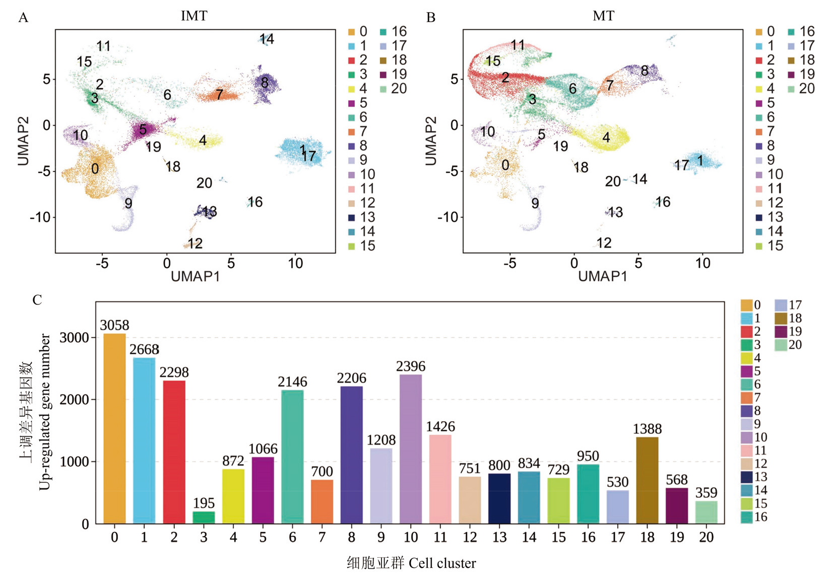
图1
鸭睾丸图谱构建和亚群上调差异基因分析"
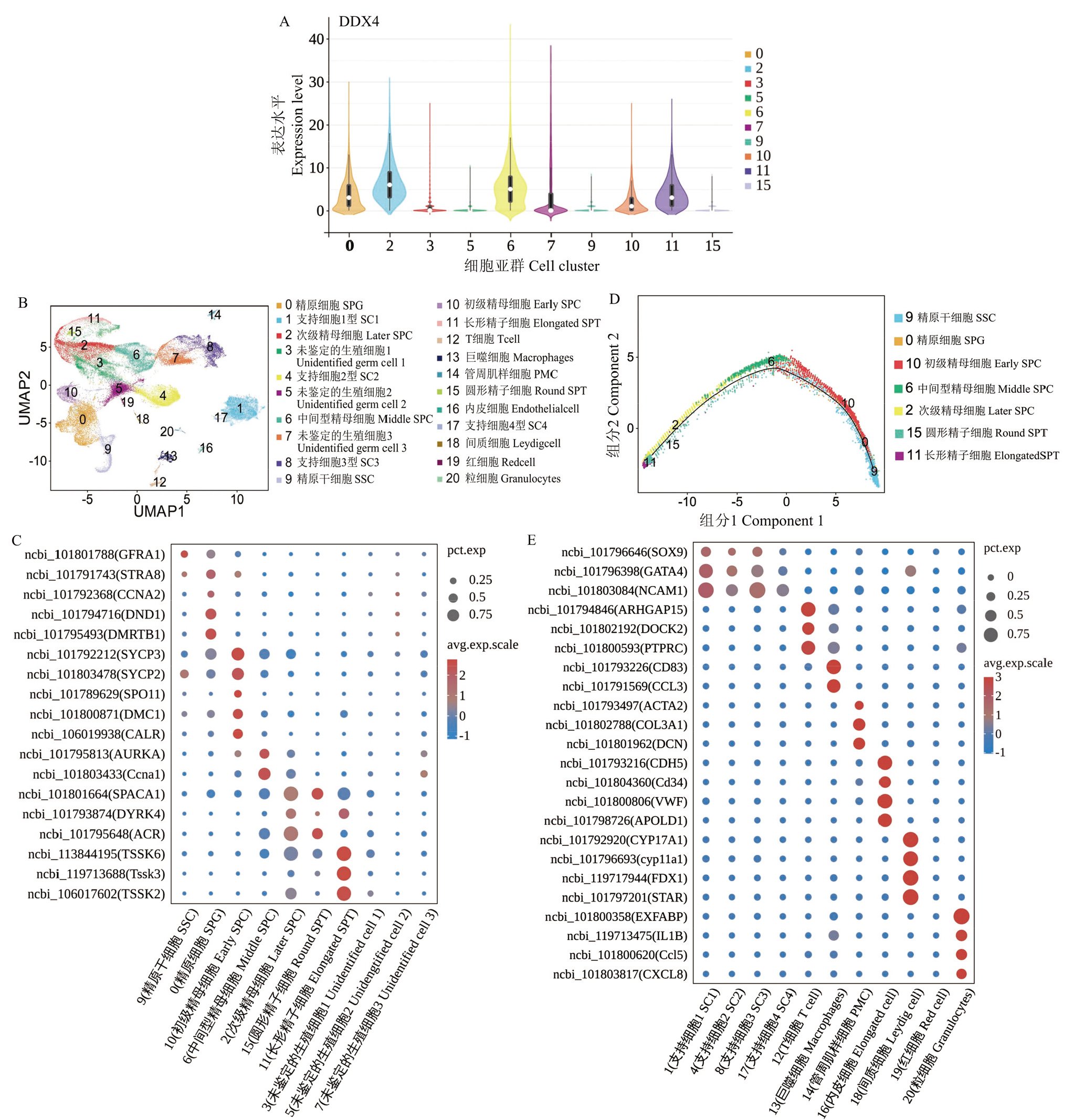
图2
鸭睾丸细胞亚群标记基因识别 A:DDX4在几个生殖细胞亚群中的表达小提琴图;B:各细胞亚群的标记基因识别UMAP图;C:各生殖细胞亚群标记基因表达气泡图;D:鉴定的7个生殖细胞亚群发育轨迹图;E:各体细胞亚群标记基因表达气泡图"
表2
不同发育时期睾丸不同亚群细胞数和细胞比率差异"
| 类群 Cluster | 细胞类型 Cell type | 未成熟睾丸组 Immature testis, IMT | 成熟睾丸组 Mature testis, MT | ||
|---|---|---|---|---|---|
| 细胞数 Cell count | 细胞比率 Cell ratio (%) | 细胞数 Cell count | 细胞比率 Cell ratio (%) | ||
| 9 | 精原干细胞 Spermatogonial stem cell | 1880 | 6.77 | 1070 | 3.97 |
| 0 | 精原细胞 Spermatogonia | 4878 | 17.57 | 1599 | 5.93 |
| 10 | 初级精母细胞 Early spermatocytes | 1383 | 4.98 | 549 | 2.04 |
| 6 | 中间型精母细胞 Middle spermatocytes | 444 | 1.60 | 3582 | 13.29 |
| 2 | 次级精母细胞 Later spermatocytes | 44 | 0.16 | 5830 | 21.64 |
| 15 | 圆形精子细胞 Round spermatids | 12 | 0.04 | 339 | 1.26 |
| 11 | 长形精子细胞 Elongated spermatids | 7 | 0.03 | 1497 | 5.56 |
| 3 | 未鉴定的生殖细胞1 Unidentified cell 1 | 2266 | 8.16 | 3199 | 11.87 |
| 5 | 未鉴定的生殖细胞2 Unidentified cell2 | 3652 | 13.16 | 493 | 1.83 |
| 7 | 未鉴定的生殖细胞3 Unidentified cell 3 | 2765 | 9.96 | 1132 | 4.20 |
| 8 | 支持细胞1型 Sertoli cell 3 | 4492 | 16.18 | 1832 | 6.80 |
| 4 | 支持细胞2型 Sertoli cell 2 | 1305 | 4.70 | 3902 | 14.48 |
| 1 | 支持细胞3型 Sertoli cell 1 | 2561 | 9.23 | 1157 | 4.29 |
| 17 | 支持细胞4型 Sertoli cell 4 | 1 | 0 | 194 | 0.72 |
| 12 | T细胞T cell | 737 | 2.66 | 58 | 0.22 |
| 13 | 巨噬细胞Macrophages | 603 | 2.17 | 78 | 0.29 |
| 14 | 管周肌样细胞 Peritubular myoid cell | 333 | 1.20 | 160 | 0.59 |
| 16 | 内皮细胞Endothelial cell | 123 | 0.44 | 121 | 0.45 |
| 18 | 间质细胞Leydig cell | 98 | 0.35 | 80 | 0.30 |
| 19 | 红细胞Red cell | 107 | 0.39 | 22 | 0.08 |
| 20 | 粒细胞Granulocyte | 65 | 0.23 | 52 | 0.19 |
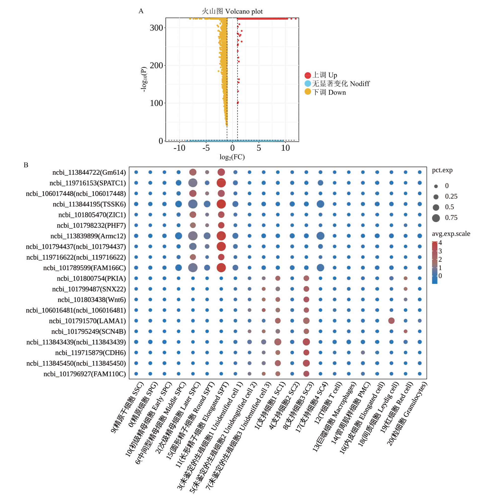
图3
不同发育时期鸭睾丸细胞差异基因分析 A:IMT和MT组间差异基因火山图;B:TOP20差异基因在各睾丸细胞亚群中的表达气泡图"
表3
不同发育时期鸭睾丸细胞TOP20差异基因列表"
| 基因ID Gene ID | 基因名称 Gene name | 差异倍数 log2FC | 描述 Description | 功能 Function |
|---|---|---|---|---|
| ncbi_113844722 | Gm614 | 11.98 | 非典型蛋白CXorf65同源异构体X2 Uncharacterized protein CXorf65 homolog isoform X2 | - |
| ncbi_119716153 | SPATC1 | 11.15 | 精子发生和中心体相关1 Spermatogenesis and centriole associated 1 | .预测能够实现r-微管蛋白结合活性 It is predicted to be capable of r-tubulin binding activity |
| ncbi_106017448 | ncbi_106017448 | 10.38 | - | - |
| ncbi_113844195 | TSSK6 | 10.34 | 睾丸特异性丝氨酸/苏氨酸蛋白激酶6 Ttestis-specific serine/threonine-protein kinase 6 | 与雄性不育和精子形态异常有关 It is associated with male sterility and abnormal sperm morphology |
| ncbi-101805470 | ZIC1 | 10.14 | 锌指蛋白ZIC 1 Zinc finger protein ZIC 1 | 在中枢神经系统器官发生的早期阶段以及脊髓背侧发育和小脑成熟过程中起着重要作用 It plays an important role in the early stages of central nervous system organogenesis, as well as in dorsal spinal cord development and cerebellar maturation |
| ncbi_101798232 | PHF7 | 9.98 | PHD手指蛋白7 PHD finger protein 7 | 在支持细胞中表达,与精子发生的转录调控有关 It is expressed in Sertoli cells and is associated with the transcriptional regulation of spermatogenesis |
| ncbi_113839899 | Armc12 | 9.98 | 含犰狳重复序列蛋白12异构体X2 Armadillo repeat-containing protein 12 isoform X2 | 参与细胞生长和精子线粒体鞘组装的正向调节,与生精失败有关 It is involved in the positive regulation of cell growth and sperm mitochondrial sheath assembly, and is associated with spermatogenic failure |
| ncbi_101794437 | ncbi_101794437 | 9.9 | - | - |
| ncbi_119716622 | ncbi_119716622 | 9.77 | - | - |
| ncbi_101789599 | FAM166C | 9.74 | UPF0573蛋白C2orf70同源物 UPF0573 protein C2orf70 homolog | 位于轴丝微管内,预测与有鞭毛的精子运动有关 It is located within the axonemal microtubules and predicted to be associated with flagellated sperm motility |
| ncbi_101800754 | PKIA | -5.11 | cAMP依赖性蛋白激酶抑制剂α cAMP-dependent protein kinase inhibitor alpha | 与PKA的Cα和Cβ催化亚基相互作用并抑制其活性 It interacts with and inhibits the activity of the Cα and Cβ catalytic subunits of PKA |
| ncbi_101799487 | SNX22 | -5.16 | 排序连接蛋白22 Sorting nexin-22 | 可能在细胞内运输中发挥作用 It may play a role in intracellular transport |
| ncbi_101803438 | Wnt6 | -5.84 | 蛋白Wnt-6亚型X1 Protein Wnt-6 isoform X1 | 参与控制未分化精原干细胞的增殖 It is involved in controlling the proliferation of undifferentiated spermatogonial stem cells |
| ncbi_106016481 | ncbi_106016481 | -5.87 | - | - |
| ncbi_101791570 | LAMA1 | -6.19 | 层粘连蛋白亚基α-1 Llaminin subunit alpha-1 | 通过高亲和力受体与细胞结合,被认为通过与其他细胞外基质成分相互作用,在胚胎发育过程中介导细胞附着、迁移和组织到组织中 It binds to cells via high-affinity receptors and is thought to mediate cell attachment, migration, and organization into tissues during embryonic development by interacting with other extracellular matrix components |
| ncbi_101795249 | SCN4B | -6.67 | X1钠通道亚基β-4亚型X1 Sodium channel subunit beta-4 isoform | 调节通道门控动力学 It regulates channel gating dynamics |
| ncbi_113843439JIGNY | ncbi_113843439 | -6.96 | 未鉴定蛋白LOC113843439 Uncharacterized protein LOC113843439 | - |
| ncbi_119715879 | CDH6 | -6.99 | 钙粘蛋白-6 Cadherin-6 | 在细胞分化和形态发生中起关键作用 It plays a critical role in cell differentiation and morphogenesis |
| ncbi_113845450 | ncbi_113845450 | -7.37 | - | - |
| ncbi_101796927 | FAM110C | -7.96 | FAM110C蛋白 Protein FAM110C | 激活a-微管蛋白活性。参与细胞迁移的正向调控 It activates α-tubulin activity and participates in the positive regulation of cell migration |

图4
不同发育时期鸭睾丸细胞差异基因功能分析 A:IMT和MT组间差异基因显著性GO分析;B:IMT和MT组间差异基因显著性KEGG分析;C:内质网中蛋白质加工信号通路差异基因的蛋白-蛋白网络互作图;D:细胞衰老信号通路差异基因的蛋白-蛋白网络互作图"
| [1] |
马荆鄂, 熊信威, 周敏, 吴斯琪, 韩甜, 饶友生, 王樟凤, 许继国. 基于垂体全长转录组测序分析鸡睾丸性状相关基因 中国农业科学, 2024, 57(20): 4130-4147. doi: 10.3864/j.issn.0578-1752.2024.20.017
doi: 10.3864/j.issn.0578-1752.2024.20.017 |
|
doi: 10.3864/j.issn.0578-1752.2024.20.017 |
|
| [2] |
卜治文, 陆璐洋, 白顺, 康振龙, 韩圣林, 叶岚. 精子发生转录调控机制的研究进展. 中国细胞生物学学报, 2024, 46(4): 595-606.
|
|
|
|
| [3] |
doi: 10.1038/s41422-018-0074-y pmid: 30061742 |
| [4] |
doi: S1934-5909(18)30392-8 pmid: 30174296 |
| [5] |
doi: 10.1038/sdata.2018.192 |
| [6] |
doi: S1534-5807(18)30636-1 pmid: 30146481 |
| [7] |
doi: 10.1093/humrep/deac245 |
| [8] |
doi: 10.7150/ijbs.82191 pmid: 37151874 |
| [9] |
高源. 安格斯牛睾丸组织非编码RNA鉴定及单细胞转录图谱绘制[D]. 杨凌: 西北农林科技大学, 2021.
|
|
|
|
| [10] |
doi: 10.3390/ijms24097982 |
| [11] |
doi: 10.1096/fsb2.v35.2 |
| [12] |
|
| [13] |
doi: 10.1186/s40104-021-00638-3 pmid: 34872612 |
| [14] |
doi: 10.3390/ijms25189786 |
| [15] |
doi: 10.1016/j.celrep.2020.03.055 |
| [16] |
doi: 10.1095/biolreprod.111.091629 pmid: 21653890 |
| [17] |
|
| [18] |
doi: S2211-1247(19)30063-4 pmid: 30726734 |
| [19] |
doi: 10.3390/cells11243978 |
| [20] |
doi: 10.1096/fsb2.v36.6 |
| [21] |
doi: 10.1371/journal.pgen.1007810 |
| [22] |
doi: 10.1080/14737159.2021.1924060 |
| [23] |
doi: 10.3389/fimmu.2021.723908 |
| [24] |
陶志云, 徐文娟, 穆金泉, 朱春红, 宋卫涛, 刘宏祥, 章双杰, 王志成, 李慧芳. 金定鸭睾丸生长发育规律的初步研究. 畜牧与兽医, 2025, 57(8): 1-6.
|
|
|
|
| [25] |
|
| [26] |
doi: 10.3390/genes13061077 |
| [27] |
|
| [28] |
doi: 10.1016/j.devcel.2022.04.004 pmid: 35504286 |
| [29] |
doi: 10.1126/science.ade8873 pmid: 36758105 |
| [30] |
doi: 10.1007/s12031-020-01620-w pmid: 32642801 |
| [31] |
doi: 10.3892/ol |
| [32] |
|
| [33] |
doi: 10.1111/ejb.2010.277.issue-3 |
| [34] |
doi: 10.1371/journal.pbio.0050105 |
| [35] |
doi: 10.1093/nar/gkn362 pmid: 18566005 |
| [36] |
|
| [37] |
doi: 10.1242/dev.122.1.53 pmid: 8565853 |
| [38] |
doi: 10.1006/dbio.1996.0007 |
| [39] |
pmid: 9041194 |
| [40] |
doi: 10.1038/3855 |
| [41] |
doi: 10.1371/journal.pone.0047862 |
| [42] |
doi: 10.1038/s41467-020-19414-4 pmid: 33173058 |
| [43] |
doi: 10.1016/j.mce.2021.111179 |
| [44] |
doi: 10.1016/S0167-4838(00)00159-X |
| [45] |
doi: 10.3389/fmicb.2022.963678 |
| [46] |
doi: 10.3390/ijms24054824 |
| [47] |
doi: 10.1016/j.ecoenv.2022.113527 |
| [48] |
苏杰. 出生后湖羊睾丸发育图谱、组织结构及X染色体剂量补偿研究[D]. 呼和浩特: 内蒙古农业大学, 2022.
|
|
|
|
| [49] |
doi: 10.1210/er.2018-00140 |
| [50] |
|
| [51] |
doi: 10.1242/jcs.047225 |
| [52] |
|
| [53] |
|
| [54] |
doi: 10.1136/jmedgenet-2021-108137 |
| [55] |
|
| [56] |
doi: 10.1186/1471-2164-11-211 pmid: 20350334 |
| [57] |
|
| [58] |
doi: 10.1074/jbc.272.32.20021 |
| [59] |
|
| [60] |
doi: 10.1016/j.yexcr.2021.112511 |
| [61] |
|
| [62] |
|
| [63] |
doi: 10.1007/978-1-4614-4015-4_7 pmid: 22872478 |
| [64] |
doi: 10.1038/s41568-020-00312-2 pmid: 33214692 |
| [65] |
doi: 10.1038/35080081 pmid: 11433366 |
| [66] |
doi: 10.1002/jemt.20787 pmid: 19941292 |
| [67] |
doi: 10.1186/1477-7827-7-12 |
| [68] |
doi: 10.1016/j.bbagen.2019.06.005 |
| [69] |
|
| [70] |
doi: 10.1093/jxb/eraa167 |
| [71] |
doi: 10.1586/14737140.8.2.207 pmid: 18279062 |
| [72] |
|
| [73] |
doi: 10.3389/fphar.2021.757120 |
| [74] |
doi: 10.1097/BS9.0000000000000009 pmid: 35402801 |
| [75] |
doi: 10.3389/fcell.2015.00016 pmid: 25914884 |
| [76] |
doi: 10.1038/nrc.2016.138 pmid: 28127048 |
| [77] |
doi: 10.1093/toxsci/kfx209 |
| [1] | 陈亚茹, 王磊, 付明, 黄涛, 张昊, 梁振华, 皮劲松, 吴艳. USP18抑制GPX4泛素化降解调控蛋鸭卵巢颗粒细胞铁死亡的分子机制[J]. 中国农业科学, 2026, 59(5): 1128-1140. |
| [2] | 刘宏祥, 张雪萍, 王逸飞, 王志成, 顾昊天, 宋卫涛, 陶志云, 徐文娟, 章双杰, 卢立志, 李慧芳, 朱春红. 金定鸭产蛋数性状的全基因组关联研究[J]. 中国农业科学, 2025, 58(15): 3145-3158. |
| [3] | 吴永保, 唐静, 曹俊婷, 王岐蒙, 谢明, 周正奎, 侯水生, 闻治国. 低能量低蛋白饲粮中添加蛋氨酸对育肥期北京鸭生长性能、屠宰性能和血浆生化指标的影响[J]. 中国农业科学, 2025, 58(12): 2475-2486. |
| [4] | 马荆鄂, 熊信威, 周敏, 吴斯琪, 韩甜, 饶友生, 王樟凤, 许继国. 基于垂体全长转录组测序分析鸡睾丸性状相关基因[J]. 中国农业科学, 2024, 57(20): 4130-4144. |
| [5] | 孙艳发, 吴琼, 林如龙, 陈红萍, 甘秋云, 沈玥, 王亚茹, 薛鹏飞, 陈飞帆, 刘健涛, 周陈鑫, 兰诗诗, 潘浩哲, 邓凡, 岳稳, 江宵兵, 李焰. 龙岩山麻鸭蛋品质性状的全基因组关联研究[J]. 中国农业科学, 2023, 56(3): 572-586. |
| [6] | 吴世豪, 黄天然, 黄明. HS-SPME-GC-MS技术结合电子鼻分析热处理对南京盐水鸭高温蒸煮味的影响[J]. 中国农业科学, 2023, 56(17): 3435-3451. |
| [7] | 吴艳,张昊,梁振华,潘爱銮,申杰,蒲跃进,黄涛,皮劲松,杜金平. circ-13267通过let-7-19/ERBB4通路调控蛋鸭卵泡颗粒细胞凋亡[J]. 中国农业科学, 2022, 55(8): 1657-1666. |
| [8] | 张亚男,金永燕,庄智威,王爽,夏伟光,阮栋,陈伟,郑春田. 鸡蛋与鸭蛋的蛋壳力学特性、超微结构及蛋壳组分的比较[J]. 中国农业科学, 2022, 55(24): 4957-4968. |
| [9] | 张卫东,郑玉杰,葛伟,张月朗,李芳,王昕. 单细胞测序对绒山羊毛乳头细胞的鉴定[J]. 中国农业科学, 2022, 55(12): 2436-2446. |
| [10] | 赵冬敏,黄欣梅,章丽娇,刘青涛,杨婧,韩凯凯,刘宇卓,李银. 坦布苏病毒感染诱导雏鸭体内未折叠蛋白反应[J]. 中国农业科学, 2021, 54(4): 855-863. |
| [11] | 刘娇,陈志敏,郑爱娟,刘国华,蔡辉益,常文环. 葡萄糖氧化酶对大肠杆菌攻毒肉鸭生长性能、免疫功能及肠道健康的影响[J]. 中国农业科学, 2021, 54(22): 4917-4930. |
| [12] | 岳盈肖,何近刚,赵江丽,闫子茹,程玉豆,武肖琦,王永霞,关军锋. 窖藏和冷藏条件下鸭梨挥发性物质及其相关基因表达分析[J]. 中国农业科学, 2021, 54(21): 4635-4649. |
| [13] | 王岭,才羿,王桂超,王迪,盛云燕. 甜瓜SLAF图谱构建及果实相关性状QTL分析[J]. 中国农业科学, 2021, 54(19): 4196-4206. |
| [14] | 柳艳霞,王振宇,郑晓春,朱瑶迪,陈丽,张德权. 基于品质指标预测北京烤鸭的中心温度[J]. 中国农业科学, 2020, 53(8): 1655-1663. |
| [15] | 崔家杰,谢强,翟双双,龚涛,朱勇文,杨琳,王文策. 光照强度对樱桃谷肉鸭c-fos、生物钟基因表达 及褪黑激素的影响[J]. 中国农业科学, 2020, 53(4): 848-856. |
|
||