


中国农业科学 ›› 2022, Vol. 55 ›› Issue (12): 2436-2446.doi: 10.3864/j.issn.0578-1752.2022.12.014
张卫东( ),郑玉杰,葛伟,张月朗,李芳,王昕()
收稿日期:2021-04-30
接受日期:2021-10-29
出版日期:2022-06-16
发布日期:2022-06-23
通讯作者:
王昕
作者简介:张卫东,E-mail: 基金资助:
ZHANG WeiDong(),ZHENG YuJie,GE Wei,ZHANG YueLang,LI Fang,WANG Xin()
Received:2021-04-30
Accepted:2021-10-29
Online:2022-06-16
Published:2022-06-23
Contact:
Xin WANG
摘要:
【目的】 基于单细胞测序技术探究绒山羊毛乳头细胞标记基因,优化毛乳头细胞体外鉴定的方法,为研究绒山羊毛囊发生发育提供良好的细胞模型。【方法】 运用Seurat单细胞分析法分析陕北白绒山羊胚胎期60、90、120 d皮肤组织的单细胞测序数据。原始数据经质控、过滤、标准化后,采用UMAP方法进行数据降维与聚类分析。通过对比已报道的毛囊相关标记基因,获得完整绒山羊毛囊细胞类群谱系信息。通过基因差异表达分析,筛选绒山羊毛乳头细胞特异性标记基因。利用免疫荧光试验鉴定标记蛋白在绒山羊皮肤组织中的表达与分布情况,进一步筛选毛乳头区域特异性表达蛋白。体视镜下机械分离完整绒山羊毛囊,采用酶消化法分离绒山羊毛乳头区域并在体外培养至细胞分离,最终通过差速贴壁法纯化细胞。待毛乳头细胞纯度较高时,利用免疫荧光试验验证候选标记蛋白在分离细胞中的表达情况。【结果】 从单细胞水平分析了绒山羊毛囊发生过程中涉及的关键细胞转录信息,成功获得绒山羊皮肤中的17个主要细胞类群信息,鉴定出包括真皮细胞谱系、表皮细胞谱系、毛乳头细胞、毛干细胞、内根鞘细胞等绒山羊皮肤结构关键细胞类群,以及周皮细胞、巨噬细胞、肌肉细胞等其他功能细胞类群。筛选获得毛乳头细胞特异性基因427个,包括SOX2、FGF7、APOD、BMP3、HHIP、HEY2和SPON1等,这些基因在绒山羊毛乳头细胞中的表达丰度远高于其他细胞类型,被认为是毛乳头细胞特异性标记基因。组织免疫荧光试验进一步证明SOX2、FGF7与APOD等蛋白在绒山羊毛乳头区域内特异性表达,可用于皮肤组织中绒山羊毛乳头细胞的定位。此外,本研究在成功分离单根绒山羊次级毛囊的基础上,实现了绒山羊毛乳头区域的贴壁培养,成功观测到大量细胞从毛乳头区域逐步迁移分离的过程。细胞免疫荧光试验结果显示SOX2、FGF7与APOD均在绒山羊毛乳头细胞中表达,且SOX2阳性细胞约占毛乳头细胞总量的76%左右,而FGF7和APOD阳性细胞则占98%以上。结合绒山羊皮肤组织免疫荧光定位结果,SOX2、FGF7与APOD等标记可用于鉴定体外分离培养的绒山羊毛乳头细胞。【结论】 利用单细胞测序技术描述了绒山羊主要皮肤细胞的转录组信息,筛选出毛乳头细胞特异性表达基因。经免疫荧光试验证实,单细胞测序鉴定标记基因的方法简单高效,且具备较高准确率。筛选的SOX2、FGF7与APOD不仅为绒山羊毛乳头细胞体内定位提供了有效的标记,而且为多标记鉴定毛乳头细胞提供可能,更为进一步研究毛囊发育相关基因的功能及调控机制奠定重要基础。
张卫东,郑玉杰,葛伟,张月朗,李芳,王昕. 单细胞测序对绒山羊毛乳头细胞的鉴定[J]. 中国农业科学, 2022, 55(12): 2436-2446.
ZHANG WeiDong,ZHENG YuJie,GE Wei,ZHANG YueLang,LI Fang,WANG Xin. Identification of Cashmere Dermal Papilla Cells Based on Single- Cell RNA Sequencing Technology[J]. Scientia Agricultura Sinica, 2022, 55(12): 2436-2446.
表1
用于细胞类群鉴定的标记基因"
| 类群ID Cluster ID | 分子标记 Marker | 细胞类型 Cell type | 参考文献 Reference |
|---|---|---|---|
| 1, 2, 3, 5, 6 | LUM, COL1A1 | Dermal | [ |
| 0,7 | KRT14, KRT17 | Epithelium | [ |
| 4,11 | MSX1, LHX2 | Hair shaft | [ |
| 8 | KRT71, KRT27 | IRS | [ |
| 9 | KRT14, KRT15 | Epithelial | [ |
| 10 | KDR, PECAM1 | Endothelial | [ |
| 12 | SOX2, SOX18 | DP | [ |
| 13 | TPM2, ACTA2 | Pericyte | [ |
| 14, 15 | ALF1, RGSI | Macrophage | [ |
| 16 | CNMD, ARSI | Muscle | [ |

图1
UMAP聚类的主要细胞分群 1、2、3、5、6为真皮细胞;0、7为表皮细胞;4、11为毛干细胞;8为内根鞘细胞;9为上皮细胞;10为内皮细胞;12为毛乳头细胞;13为周皮细胞;14、15为巨噬细胞;16为肌肉细胞"
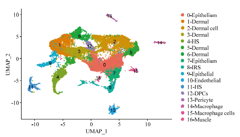
表2
DPCs特异性标记基因(前20)"
| 基因名称 Gene symbol | P | 平均log2倍数 Avg_log2 FC | 表达百分比1 Pct.1 | 表达百分比2 Pct.2 | 校正P值 P_val_adj |
|---|---|---|---|---|---|
| APOD | 0 | 2.141965302 | 0.548 | 0.028 | 0 |
| RSPO2 | 0 | 1.890455415 | 0.350 | 0.005 | 0 |
| PAPPA2 | 0 | 1.309548033 | 0.494 | 0.046 | 0 |
| BMP3 | 0 | 1.163114736 | 0.411 | 0.024 | 0 |
| SOX2 | 0 | 1.094986265 | 0.409 | 0.014 | 0 |
| PCOLCE2 | 0 | 1.080839693 | 0.272 | 0.014 | 0 |
| HEY2 | 0 | 1.040877239 | 0.442 | 0.026 | 0 |
| SPON1 | 0 | 0.996565048 | 0.558 | 0.07 | 0 |
| HHIP | 0 | 0.986923554 | 0.411 | 0.022 | 0 |
| FGF7 | 0 | 2.214535719 | 0.392 | 0.078 | 0 |
| SMOC1 | 0 | 0.952556549 | 0.57 | 0.069 | 0 |
| ITGA8 | 0 | 0.906779411 | 0.525 | 0.051 | 0 |
| SCUBE3 | 0 | 0.818127433 | 0.307 | 0.004 | 0 |
| NRG2 | 0 | 0.810698041 | 0.281 | 0.012 | 0 |
| PREX2 | 2.04E-303 | 1.000680599 | 0.584 | 0.079 | 3.42E-299 |
| FGFR1 | 4.44E-281 | 1.453693627 | 0.842 | 0.224 | 7.44E-277 |
| IGF1 | 2.89E-274 | 1.446981853 | 0.835 | 0.193 | 4.84E-270 |
| GAL | 1.30E-272 | 1.230618938 | 0.286 | 0.019 | 2.18E-268 |
| INHBA | 4.25E-269 | 1.55167208 | 0.780 | 0.188 | 7.12E-265 |
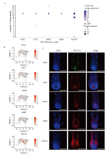
图2
DPCs特异性基因的筛选与鉴定 A:点状图展示标记基因在cluster内的分布;B:细胞散点图显示标记基因在细胞分群中的位置;C:免疫荧光验证标记基因在绒山羊毛囊中的表达位置,DAPI:细胞核染料、FITC/CY3:选用的荧光标记分子,Merge:组合图,100 μm"

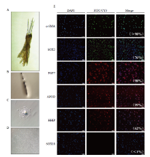
图3
绒山羊DPCs的分离过程及标记基因的鉴定 A:绒山羊次级毛囊簇;B:单根绒山羊次级毛囊;C:毛乳头区域分离的细胞;D:经纯化的毛乳头细胞;E:免疫荧光验证标记基因在毛乳头细胞中的表达,100 μm,组合图右下角为阳性细胞率(荧光阳性细胞数/总细胞数)"

| [1] |
MA S, WANG Y, ZHOU G X, DING Y, YANG Y X, WANG X L, ZHANG E P, CHEN Y L. Synchronous profiling and analysis of mRNAs and ncRNAs in the dermal papilla cells from cashmere goats. BMC Genomics, 2019, 20(1): 512. doi: 10.1186/s12864-019-5861-4.
doi: 10.1186/s12864-019-5861-4 |
| [2] |
PAUS R, MÜLLER-RÖVER S, VAN DER VEEN C, MAURER M, EICHMÜLLER S, LING G, HOFMANN U, FOITZIK K, MECKLENBURG L, HANDJISKI B. A comprehensive guide for the recognition and classification of distinct stages of hair follicle morphogenesis. The Journal of Investigative Dermatology, 1999, 113(4): 523-532. doi: 10.1046/j.1523-1747.1999.00740.x.
doi: 10.1046/j.1523-1747.1999.00740.x. |
| [3] |
STENN K S, PAUS R. Controls of hair follicle cycling. Physiological Reviews, 2001, 81(1): 449-494. doi: 10.1152/physrev.2001.81.1.449.
doi: 10.1152/physrev.2001.81.1.449 |
| [4] |
HARSHUK-SHABSO S, DRESSLER H, NIEHRS C, AAMAR E, ENSHELL-SEIJFFERS D. Fgf and Wnt signaling interaction in the mesenchymal niche regulates the murine hair cycle clock. Nature Communications, 2020, 11(1): 5114. doi: 10.1038/s41467-020-18643-x.
doi: 10.1038/s41467-020-18643-x |
| [5] |
NAMEKATA M, YAMAMOTO M, GOITSUKA R. Nuclear localization of Meis1 in dermal papilla promotes hair matrix cell proliferation in the anagen phase of hair cycle. Biochemical and Biophysical Research Communications, 2019, 519(4): 727-733. doi: 10.1016/j.bbrc.2019.09.060.
doi: 10.1016/j.bbrc.2019.09.060 |
| [6] |
CHI W, WU E, MORGAN B A. Dermal papilla cell number specifies hair size, shape and cycling and its reduction causes follicular decline. Development (Cambridge, England), 2013, 140(8): 1676-1683. doi: 10.1242/dev.090662.
doi: 10.1242/dev.090662 |
| [7] |
DRISKELL R R, GIANGRECO A, JENSEN K B, MULDER K W, WATT F M. Sox2-positive dermal papilla cells specify hair follicle type in mammalian epidermis. Development (Cambridge, England), 2009, 136(16): 2815-2823. doi: 10.1242/dev.038620.
doi: 10.1242/dev.038620 |
| [8] |
CLAVEL C, GRISANTI L, ZEMLA R, REZZA A, BARROS R, SENNETT R, MAZLOOM A R, CHUNG C Y, CAI X, CAI C L, PEVNY L, NICOLIS S, MA'AYAN A, RENDL M. Sox 2 in the dermal papilla niche controls hair growth by fine-tuning BMP signaling in differentiating hair shaft progenitors. Developmental Cell, 2012, 23(5): 981-994. doi: 10.1016/j.devcel.2012.10.013.
doi: 10.1016/j.devcel.2012.10.013 |
| [9] |
REYNOLDS A J, CHAPONNIER C, JAHODA C A, GABBIANI G. A quantitative study of the differential expression of alpha-smooth muscle actin in cell populations of follicular and non-follicular origin. The Journal of Investigative Dermatology, 1993, 101(4): 577-583. doi: 10.1111/1523-1747.ep12366032.
doi: 10.1111/1523-1747.ep12366032 |
| [10] |
PENNISI D, GARDNER J, CHAMBERS D, HOSKING B, PETERS J, MUSCAT G, ABBOTT C, KOOPMAN P. Mutations in Sox18 underlie cardiovascular and hair follicle defects in ragged mice. Nature Genetics, 2000, 24(4): 434-437. doi: 10.1038/74301.
doi: 10.1038/74301 |
| [11] |
ITO Y, HAMAZAKI T S, OHNUMA K, TAMAKI K, ASASHIMA M, OKOCHI H. Isolation of murine hair-inducing cells using the cell surface marker prominin-1/CD133. The Journal of Investigative Dermatology, 2007, 127(5): 1052-1060. doi: 10.1038/sj.jid.5700665.
doi: 10.1038/sj.jid.5700665 |
| [12] |
VILLANI R, HODGSON S, LEGRAND J, GREANEY J, WONG H Y, PICHOL-THIEVEND C, ADOLPHE C, WAINWIGHT B, FRANCOIS M, KHOSROTEHRANI K. Dominant-negative Sox18 function inhibits dermal papilla maturation and differentiation in all murine hair types. Development (Cambridge, England), 2017, 144(10): 1887-1895. doi: 10.1242/dev.143917.
doi: 10.1242/dev.143917 |
| [13] |
ZHOU L, XU M, YANG Y, YANG K, WICKETT R R, ANDL T, MILLAR S E, ZHANG Y. Activation of β-catenin signaling in CD133-positive dermal papilla cells drives postnatal hair growth. PLoS One, 2016, 11(7): e0160425. doi: 10.1371/journal.pone.0160425.
doi: 10.1371/journal.pone.0160425 |
| [14] |
DRISKELL R R, JUNEJA V R, CONNELLY J T, KRETZSCHMAR K, TAN D W-M, WATT F M. Clonal growth of dermal papilla cells in hydrogels reveals intrinsic differences between Sox2- positive and -negative cells in vitro and in vivo. Journal of Investigative Dermatology, 2012, 132(4), 1084-1093. doi: 10.1038/jid.2011.428.
doi: 10.1038/jid.2011.428 |
| [15] |
TSAI S Y, CLAVEL C, KIM S, ANG Y S, GRISANTI L, LEE D F, KELLEY K, RENDL M. Oct4 and klf4 reprogram dermal papilla cells into induced pluripotent stem cells. Stem Cells, 2010, 28(2):221-228. doi: 10.1002/stem.281.
doi: 10.1002/stem.281 |
| [16] |
WANG Y, MACK J A, MAYTIN E V. CD 44 inhibits α-SMA gene expression via a novel G-actin/MRTF-mediated pathway that intersects with TGFβR/p38MAPK signaling in murine skin fibroblasts. The Journal of Biological Chemistry, 2019, 294(34): 12779-12794. doi: 10.1074/jbc.ra119.007834.
doi: 10.1074/jbc.ra119.007834 |
| [17] |
SOLDANO S, MONTAGNA P, BRIZZOLARAR. AB 0238 Effects of endothelin A/B receptor antagonist (bosentan) on alpha-smooth muscle actin (α-SMA) and extracellular matrix protein synthesis in primary cultures of systemic sclerosis skin fibroblasts. Annals of the Rheumatic Diseases, 2013, 71: 651. doi: 10.4081/reumatismo.2012.326.
doi: 10.4081/reumatismo.2012.326 |
| [18] |
HAM S A, HWANG J S, YOO T, LEE W J, PAEK K S, OH J W, PARK C K, KIM J H, DO J T, KIM J H, SFO H G. Ligand-activated PPARδ upregulates α-smooth muscle actin expression in human dermal fibroblasts: a potential role for PPARδ in wound healing. Journal of Dermatological Science, 2015, 80(3): 186-195. doi: 10.1016/j.jdermsci.2015.10.005.
doi: 10.1016/j.jdermsci.2015.10.005 |
| [19] |
HE X L, CHAO Y, ZHOU G X, CHEN Y L. Fibroblast growth factor 5-short (FGF5s) inhibits the activity of FGF5 in primary and secondary hair follicle dermal papilla cells of cashmere goats. Gene, 2016, 575(2 pt 2): 393-398. doi: 10.1016/j.gene.2015.09.034.
doi: 10.1016/j.gene.2015.09.034 |
| [20] |
ZHU B, XU T, YUAN J, GUO X, LIU D. Transcriptome sequencing reveals differences between primary and secondary hair follicle-derived dermal papilla cells of the cashmere goat (Capra hircus). 2013, 46(3):104-111. doi: 10.1371/journal.pone.0076282.
doi: 10.1371/journal.pone.0076282 |
| [21] |
MA S, ZHOU G, CHEN Y. Effects of all-trans retinoic acid on goat dermal papilla cells cultured in vitro. Electronic Journal of Biotechnology, 2018, 34: 43-50. doi: 10.1016/j.ejbt.2018.05.004.
doi: 10.1016/j.ejbt.2018.05.004 |
| [22] |
JAHO DA C A, REYNOLDS A J, CHAPONNIER C, FORESTER J. C, GABBIANI G. Smooth muscle alpha-actin is a marker for hair follicle dermis in vivo and in vitro. Journal of Cell Science, 1991, 99 (Pt 3)(2):627. doi: 10.1242/jcs.99.3.627.
doi: 10.1242/jcs.99.3.627 |
| [23] |
ZHU B, GUO Z L, JIN M Z, BAI Y J, YANG W L, HUI L H. Establishment of dermal sheath cell line from Cashmere goat and characterizing cytokeratin 13 as its novel biomarker. Biotechnology Letters, 2018, 40(5): 765-772. doi: 10.1007/s10529-018-2532-5.
doi: 10.1007/s10529-018-2532-5 |
| [24] |
JOOST S, ZEISEL A, JACOB T, SUN X, LA MANNO G, LÖNNERBERG P, LINNARSSON S, KASPER M. Single-cell transcriptomics reveals that differentiation and spatial signatures shape epidermal and hair follicle heterogeneity. Cell Systems, 2016, 3(3): 221-237.e9. doi: 10.1016/j.cels.2016.08.010.
doi: 10.1016/j.cels.2016.08.010 |
| [25] |
JAITIN D A, KENIGSBERG E, KEREN-SHAUL H, ELEFANT N, PAUL F, ZARETSKY I, MILDNER A, COHEN N, JUNG S, TANAY A, AMIT I. Massively parallel single-cell RNA-seq for marker-free decomposition of tissues into cell types. Science, 2014, 343(6172): 776-779. doi: 10.1126/science.1247651.
doi: 10.1126/science.1247651 |
| [26] |
GE W, TAN S J, WANG S H, LI L, SUN X F, SHEN W, WANG X. Single-cell transcriptome profiling reveals dermal and epithelial cell fate decisions during embryonic hair follicle development. Theranostics, 2020, 10(17): 7581-7598. doi: 10.7150/thno.44306.
doi: 10.7150/thno.44306 |
| [27] |
GUPTA K, LEVINSOHN J, LINDERMAN G, CHEN D, SUN T Y, DONG D, TAKETO M M, BOSENBERG M, KLUGER Y, CHOATE K. Single-cell analysis reveals a hair follicle dermal niche molecular differentiation trajectory that begins prior to morphogenesis. Developmental Cell, 2019, 48(1):17-31. doi: 10.1016/j.devcel.2018.11.032.
doi: 10.1016/j.devcel.2018.11.032 |
| [28] |
CHOVATIYA G, GHUWALEWALA S, WALTER L D, COSGROVE B D, TUMBAR T. High resolution single cell transcriptomics reveals heterogeneity of self‐renewing hair follicle stem cells. Experimental Dermatology, 2020, 30(4): 457-471. doi: 10.1111/exd.14262.
doi: 10.1111/exd.14262 |
| [29] |
GUPTA K, LEVINSOHN J, LINDERMAN G, CHEN D, SUN T Y, DONG D, TAKETO M M, BOSENBERG M, KLUGER Y, CHOATE K, MYUNG P. Single-cell analysis reveals a hair follicle dermal niche molecular differentiation trajectory that begins prior to morphogenesis. Developmental Cell, 2019, 48(1): 17-31.e6. doi: 10.1016/j.devcel.2018.11.032.
doi: 10.1016/j.devcel.2018.11.032 |
| [30] |
GU L H, COULOMBE P A. Keratin function in skin epithelia: a broadening palette with surprising shades. Current Opinion in Cell Biology, 2007, 19(1): 13-23. doi: 10.1016/j.ceb.2006.12.007.
doi: 10.1016/j.ceb.2006.12.007 |
| [31] |
YANG H, ADAM R C, GE Y, HUA Z L, FUCHS E. Epithelial-mesenchymal micro-niches govern stem cell lineage choices. Cell, 2017, 169(3): 483-496.e13. doi: 10.1016/j.cell.2017.03.038.
doi: 10.1016/j.cell.2017.03.038 |
| [32] |
HAREL S, CHRISTIANO A M. Keratin 71 mutations: from water dogs to woolly hair. Journal of Investigative Dermatology, 2012, 132(10):2315-2317. doi: 10.1038/jid.2012.291.
doi: 10.1038/jid.2012.291 |
| [33] |
TAI G, RANJZAD P, MARRIAGE F, REHMAN S, DENLEY H, DIXON J, MITCHELL K, DAY P J, WOOLF A S. Cytokeratin 15 marks basal epithelia in developing ureters and is upregulated in a subset of urothelial cell carcinomas. PLoS One, 2013, 8(11): e81167. doi: 10.1371/journal.pone.0081167.
doi: 10.1371/journal.pone.0081167 |
| [34] |
DETMAR M, BROWN L F, SCHÖN M P, ELICKER B M, VELASCO P, RICHARD L, FUKUMURA D, MONSKY W, CLAFFEY K P, JAIN R K. Increased microvascular density and enhanced leukocyte rolling and adhesion in the skin of VEGF transgenic mice. The Journal of Investigative Dermatology, 1998, 111(1): 1-6. doi: 10.1046/j.1523-1747.1998.00262.x.
doi: 10.1046/j.1523-1747.1998.00262.x. |
| [35] |
PAQUET-FIFIELD S, SCHLUTER H, LI A, AITKEN T, GANGATIRKAR P, BLASHKI D, KOELMEYER R, POULIOT N, PALATSIDES M, ELLIS S, et al. A role for pericytes as microenvironmental regulators of human skin tissue regeneration. Journal of Clinical Investigation, 2009, 119(9):2795-2806. doi: 10.1172/JCI38535.
doi: 10.1172/JCI38535 |
| [36] |
LEE S B, SHIM S, KIM M J, SHIN H Y, JANG W S, LEE S J, JIN Y W, LEE S S, PARK S. Identification of a distinct subpopulation of fibroblasts from murine dermis: CD73 (-) CD105(+) as potential marker of dermal fibroblasts subset with multipotency. Cell Biology International, 2016, 40(9): 1008-1016. doi: 10.1002/cbin.10623.
doi: 10.1002/cbin.10623 |
| [37] |
SARI A R, RUFAUT N W, JONES L N, SINCLAIR R D. Characterization of ovine dermal papilla cell aggregation. International Journal of Trichology, 2016, 8(3): 121-129. doi: 10.4103/0974-7753.188966.
doi: 10.4103/0974-7753.188966 |
| [38] |
SAXENA N, MOK K W, RENDL M. An updated classification of hair follicle morphogenesis. Experimental Dermatology, 2019, 28(4): 332-344. doi: 10.1111/exd.13913.
doi: 10.1111/exd.13913 |
| [39] |
RAHMANI W, ABBASI S, HAGNER A, RAHARJO E, KUMAR R, HOTTA A, MAGNESS S, METZGER D, BIERNASKIE J. Hair follicle dermal stem cells regenerate the dermal sheath, repopulate the dermal papilla, and modulate hair type. Developmental Cell, 2014, 31(5): 543-558. doi: 10.1016/j.devcel.2014.10.022.
doi: 10.1016/j.devcel.2014.10.022 |
| [40] |
MILLAR S E. Molecular mechanisms regulating hair follicle development. The Journal of Investigative Dermatology, 2002, 118(2): 216-225. doi: 10.1046/j.0022-202x.2001.01670.x.
doi: 10.1046/j.0022-202x.2001.01670.x. |
| [41] |
MESSENGER A G. The culture of dermal papilla cells from human hair follicles. British Journal of Dermatology, 1984, 110(6): 685-689. doi: 10.1111/j.1365-2133.1984.tb04705.x.
doi: 10.1111/j.1365-2133.1984.tb04705.x. |
| [42] |
WITHERS A P, JAHODA C A B, RYDER M L, OLIVER R F. Culture of wool follicle dermal papilla cells from two breeds of sheep. Archives of Dermatological Research, 1986, 279(2): 140-142. doi: 10.1007/BF00417536.
doi: 10.1007/BF00417536 |
| [43] |
GUO H, XING Y, ZHANG Y, HE L, DENG F, MA X, LI Y. Establishment of an immortalized mouse dermal papilla cell strain with optimized culture strategy. PeerJ, 2018, 6: e4306. doi: 10.7717/peerj.4306.
doi: 10.7717/peerj.4306 |
| [44] |
HIGGINS C A, RICHARDSON G D, FERDINANDO D, WESTGATE G E, JAHODA C A. Modelling the hair follicle dermal papilla using spheroid cell cultures. Experimental Dermatology, 2010, 19(6): 546-548. doi: 10.1111/j.1600-0625.2009.01007.x.
doi: 10.1111/j.1600-0625.2009.01007.x. |
| [45] |
OSADA A, KOBAYASHI K, MASUI S, HAMAZAKI T S, YASUDA K, OKOCHI H. Cloned cells from the murine dermal papilla have hair-inducing ability. Journal of Dermatological Science, 2009, 54(2): 129-131. doi: 10.1016/j.jdermsci.2008.12.002.
doi: 10.1016/j.jdermsci.2008.12.002 |
| [46] |
JAMES K, HOSKING B, GARDNER J, MUSCAT G E, KOOPMAN P. Sox 18 mutations in the ragged mouse alleles ragged-like and opossum. Genesis (New York, N Y), 2003, 36(1): 1-6. doi: 10.1002/gene.10190.
doi: 10.1002/gene.10190 |
| [47] |
SEO H S, LEE D J, CHUNG J H, LEE C H, KIM H R, KIM J E, KIM B J, JUNG M H, HA K T, JEONG H S. Hominis Placenta facilitates hair re-growth by upregulating cellular proliferation and expression of fibroblast growth factor-7. BMC Complementary and Alternative Medicine, 2016, 16(1): 187. doi: 10.1186/s12906-016-1180-3,
doi: 10.1186/s12906-016-1180-3 |
| [48] |
MESLER A L, VENIAMINOVA N A, LULL M V, WONG S Y. Hair follicle terminal differentiation is orchestrated by distinct early and late matrix progenitors. Cell Reports, 2017, 19(4): 809-821. doi: 10.1016/j.celrep.2017.03.077.
doi: 10.1016/j.celrep.2017.03.077 |
| [49] |
PLEASANTINE M, RONG M, HONG F, MARINA G, KIM PETER C W, DLUGOSZ ANDRZEJ A, CHI-CHUNG H. Sonic hedgehog- dependent activation of Gli2 is essential for embryonic hair follicle development. Genes & Development, 2007(2): 282. doi: 10.1101/gad.1038103.
doi: 10.1101/gad.1038103 |
| [50] |
WOO W M, ZHEN H H, ORO A E. Shh maintains dermal papilla identity and hair morphogenesis via a Noggin-Shh regulatory loop. Genes & Development, 2012, 26(11): 1235-1246. doi: 10.1101/gad.187401.112.
doi: 10.1101/gad.187401.112 |
| [1] | 王梦蕊, 刘淑梅, 侯丽霞, 王施慧, 吕宏君, 苏晓梅. 番茄颈腐根腐病抗性鉴定技术的建立及抗性种质资源筛选[J]. 中国农业科学, 2022, 55(4): 707-718. |
| [2] | 李龙, 李超男, 毛新国, 王景一, 景蕊莲. 作物根系表型鉴定评价方法的现状与展望[J]. 中国农业科学, 2022, 55(3): 425-437. |
| [3] | 杜金霞,李奕莎,李美霖,陈文浛,张木清. 甘蔗不同基因型对白条病抗性的评价[J]. 中国农业科学, 2022, 55(21): 4118-4130. |
| [4] | 琚铭, 苗红梅, 黄盈盈, 马琴, 王慧丽, 王翠英, 段迎辉, 韩秀花, 张海洋. 芝麻种间杂交亲和性差异及杂种生物学特征分析[J]. 中国农业科学, 2022, 55(20): 3897-3909. |
| [5] | 段灿星,曹言勇,董怀玉,夏玉生,李红,胡清玉,杨知还,王晓鸣. 玉米种质资源抗腐霉茎腐病和镰孢茎腐病精准鉴定[J]. 中国农业科学, 2022, 55(2): 265-279. |
| [6] | 孟雨,温鹏飞,丁志强,田文仲,张学品,贺利,段剑钊,刘万代,冯伟. 基于热红外图像的小麦品种抗旱性鉴定与评价[J]. 中国农业科学, 2022, 55(13): 2538-2551. |
| [7] | 徐晓,任根增,赵欣蕊,常金华,崔江慧. 中国高粱地方品种和育成品种穗部表型性状精准鉴定及综合评价[J]. 中国农业科学, 2022, 55(11): 2092-2108. |
| [8] | 刘闯,高振,姚玉新,杜远鹏. 葡萄钾离子转运基因VviHKT1;7在盐胁迫下的功能鉴定[J]. 中国农业科学, 2021, 54(9): 1952-1963. |
| [9] | 习玲, 王昱琦, 杨修, 朱微, 陈国跃, 王益, 覃鹏, 周永红, 康厚扬. 243份云南普通小麦地方品种抗条锈病鉴定及分子标记检测[J]. 中国农业科学, 2021, 54(4): 684-695. |
| [10] | 张鹏飞,史良玉,刘家鑫,李洋,吴成斌,王立贤,赵福平. 畜禽全基因组长纯合片段检测的研究进展[J]. 中国农业科学, 2021, 54(24): 5316-5326. |
| [11] | 陈豆豆, 关利平, 贺亮亮, 宋银花, 章鹏, 刘三军. 葡萄无核基因分子标记的通用性鉴定[J]. 中国农业科学, 2021, 54(22): 4880-4893. |
| [12] | 王程利,尹志远,聂嘉俊,林永辉,黄丽丽. 苹果黑腐皮壳菌CAP超家族蛋白基因鉴定及毒性功能分析[J]. 中国农业科学, 2021, 54(16): 3440-3450. |
| [13] | 郭志雄,孙冷雪,郑嘉敏,蔡灿军,王蓓,李开拓,潘腾飞,佘文琴,陈桂信,潘东明. 荔枝果皮BPox的分离纯化及其在果实成熟过程中的表达[J]. 中国农业科学, 2021, 54(16): 3502-3513. |
| [14] | 黄金凤,吕天星,王寻,王颖达,王冬梅,闫忠业,刘志. 苹果LRR-RLK基因家族鉴定和表达分析[J]. 中国农业科学, 2021, 54(14): 3097-3112. |
| [15] | 王富强,张建,温常龙,樊秀彩,张颖,孙磊,刘崇怀,姜建福. 基于KASP标记的葡萄品种鉴定[J]. 中国农业科学, 2021, 54(13): 2830-2842. |
|
||