| [1] |
BEGLEY C G, LOPEZ A F, NICOLA N A, WARREN D J, VADAS M A, SANDERSON C J, METCALF D. Purified colony-stimulating factors enhance the survival of human neutrophils and eosinophils in vitro: a rapid and sensitive microassay for colony-stimulating factors. Blood, 1986,68(1):162-166.
doi: 10.1182/blood.V68.1.162.162
|
| [2] |
CLARK S C, KAMEN R. The human hematopoietic colony- stimulating factors. Science, 1987,236(4806):1229-1237.
doi: 10.1126/science.3296190
|
| [3] |
AVALOS B R, GASSON J C, HEDVAT C, QUAN S G, BALDWIN G C, WEISBART R H, WILLIAMS R E, GOLDE D W, DIPERSIO J F. Human granulocyte colony-stimulating factor: biologic activities and receptor characterization on hematopoietic cells and small cell lung cancer cell lines. Blood, 1990,75(4):851-857.
doi: 10.1182/blood.V75.4.851.851
|
| [4] |
柯敏霞, 纪猛, 王皓, 洪丹萍, 吴月红, 齐念民. 干细胞模型研究进展及商业化应用的现状. 中国组织工程研究, 2018,22(5):766-773.
|
|
KE M X, JI M, WANG H, HONG D P, WU Y H, QI N M. Stem cell models for commercialization. Chinese Journal of Tissue Engineering Research, 2018,22(5):766-773. (in Chinese)
|
| [5] |
王聪慧, 赵帅, 张译元, 王立民, 李炜杰, 赵兴旺, 丁新平, 张银国, 周平. 电穿孔转染制备绵羊诱导多潜能干细胞条件的优化. 西北农业学报, 2015,24(9):9-15.
|
|
WANG C H, ZHAO S, ZHANG Y Y, WANG L M, LI W J, ZHAO X W, DING X P, ZHANG Y G, ZHOU P. Optimization of inducting pluripotent stem cells by electroporation in sheep. Acta Agriculturae Boreali-occidentalis Sinica, 2015,24(9):9-15. (in Chinese)
|
| [6] |
王峰, 刘灿, 潘传英. 大家畜诱导多潜能干细胞(iPSCs)研究进展. 农业生物技术学报, 2014,22(10):1286-1297.
|
|
WANG F, LIU C, PAN C Y. Progress of induced pluripotent stem cells (iPSCs) of big domestic animals. Journal of Agricultural Biotechnology, 2014,22(10):1286-1297. (in Chinese)
|
| [7] |
邰大鹏. 阿尔巴斯白绒山羊诱导性多潜能干细胞生成的研究[D]. 呼和浩特: 内蒙古大学, 2016.
|
|
TAI D P. Research on generation of arbas cashmere goat induced pluripotent stem cells[D]. Huhehaote: Inner Mongolia University, 2016. (in Chinese)
|
| [8] |
王聪慧. 绵羊诱导多能性干细胞的制作与鉴定研究[D]. 石河子:石河子大学, 2015.
|
|
WANG C H. Production and identification of sheep induced pluripotent stem cells[D]. Shihezi: Shihezi University, 2015. (in Chinese)
|
| [9] |
李海红, 周岗, 付小兵, 屈振亮, 孙同柱, 顾绍峰. 人皮肤成纤维细胞的培养和鉴定. 中国危重病急救医学, 2005(2):89-91+63.
|
|
LI H H, ZHOU G, FU X B, QU Z L, SUN T Z, GU S F. Culture and identification of human skin fibroblasts. Chinese Critical Care Medicine, 2005(2):89-91+63. (in Chinese)
|
| [10] |
CIBELLI J B, STICE S L, GOLUEKE P J, KANE J J, JERRY J, BLACKWELL C, PONCE DE LEON F A, ROBL J M. Cloned transgenic calves produced from nonquiescent fetal fibroblasts. Science, 1998,280(5367):1256-1258.
doi: 10.1126/science.280.5367.1256
|
| [11] |
黄兰兰, 石国庆, 白洁. 绵羊胚胎成纤维细胞的体外培养. 黑龙江动物繁殖, 2006,14(2):4-5.
|
|
HUANG L L, SHI G Q, BAI J. In vitro culture of sheep embryonic fibroblasts. Heilongjiang Journal of Animal Reproduction, 2006,14(2):4-5. (in Chinese)
|
| [12] |
DO B H, KANG H J, SONG J A, NGUYEN M T, PARK S, YOO J, NGUYEN A N, KWON G G, JANG J, JANG M, LEE S, SO S, SIM S, LEE K J, OSBORN M J, CHOE H. Granulocyte colony-stimulating factor (GCSF) fused with Fc Domain produced from E. coli is less effective than Polyethylene Glycol-conjugated GCSF. Scientific Reports, 2017,7(1):1-9.
doi: 10.1038/s41598-016-0028-x
|
| [13] |
HATFIELD K J, MELVE G K, BRUSERUD O. Granulocyte colony-stimulating factor alters the systemic metabolomic profile in healthy donors. Metabolomics, 2017,13(1):2.
doi: 10.1007/s11306-016-1139-x
|
| [14] |
ICHINOSE Y, HARA N, OHTA M, ASO H, CHIKAMA H, KAWASAKI M, KUBOTA I, SHIMIZU T, YAGAWA K. Recombinant granulocyte colony-stimulating factor and lipopolysaccharide maintain the phenotype of and superoxide anion generation by neutrophils. Infection and Immunity, 1990,58(6):1647-1652.
doi: 10.1128/iai.58.6.1647-1652.1990
|
| [15] |
CULLOR J S, SMITH W, FAIRLEY N, WOOD S L, DELLINGER J D, SOUZA L. Effects of human recombinant granulocyte colony stimulating factor (HR-GCSF) on the hemogram of lactating dairy cattle. Veterinary Clinical Pathology, 1990,19(1):9-12.
doi: 10.1111/vcp.1990.19.issue-1
|
| [16] |
NIKOLAY N, EVELIINA P, ANTTI K, RAISA G, ANASTASIA S, ANASTASIA S, JOHANNA M, JARI K, JARI K, RASHID G. Gender specific mechanism of synaptic impairment and its prevention by GCSF in a mouse model of ALS. Frontiers in Cellular Neuroscience, 2011,5:26.
|
| [17] |
MISHRA V V, CHOUDHARY S, SHARMA U, AGGARWAL R, AGARWAL R, GANDHI K, GORANIYA N. Effects of Granulocyte Colony-Stimulating Factor (GCSF) on persistent thin endometrium in Frozen Embryo Transfer (FET) cycles. Journal of Obstetrics and Gynaecology of India, 2016,66(Suppl 1):407-411.
doi: 10.1007/s13224-015-0775-9
|
| [18] |
安娜-玛莉亚·A, 海斯·蒲楠, 尼克·克努森, 席雅·诺曼, 爱伦·柯钦, 瓦迪姆·克赖诺夫, 莉莲·何, 彼德·C, 康宁. 经修饰的牛G-CSF多肽和其用途. CN102159230A, 2011-08-17.
|
|
HAYS P A M A, KNUDSEN N, NORMAN T, KODER A, KRAYNOV V, HO L, CANNING P C. Modified bovine g-csf polypeptides and their uses. China. CN102159230A, 2011-08-17. (in Chinese)
|
| [19] |
ZHANG Y, AXIAK-BECHTEL S, FRIEDMAN COWAN C, AMORIM J, TSURUTA K, DECLUE A E. Evaluation of immunomodulatory effect of recombinant human granulocyte-macrophage colony-stimulating factor on polymorphonuclear cell from dogs with cancer in vitro. Veterinary and Comparative Oncology, 2017,15(3):968-979.
doi: 10.1111/vco.2017.15.issue-3
|
| [20] |
张薇, 许嵩, 张治芬. M-CSF对卵泡颗粒细胞功能调节及其分子机制. 医学研究杂志, 2016,45(1):100-105.
|
|
ZHANG W, XU S, ZHANG Z F. Involvement of macrophage colony-stimulating factor (M-CSF) in the function of follicular granulosa cells and its molecular mechanism. Journal of Medical Research, 2016,45(1):100-105. (in Chinese)
|
| [21] |
王欣悦, 石田培, 赵志达, 胡文萍, 尚明玉, 张莉. 基于绵羊胚胎骨骼肌蛋白质组学的PI3K-AKT信号通路分析. 中国农业科学, 2020,53(14):2956-2963.
|
|
WANG X Y, SHI T P, ZHAO Z D, HU W P, SHANG M Y, ZHANG L. The analysis of PI3K-AKT signal pathway based on the proteomic results of sheep embryonic skeletal muscle. Scientia Agricultura Sinica, 2020,53(14):2956-2963. (in Chinese)
|
| [22] |
李婷, 马爱团, 张英杰, 刘月琴, 段春辉. RNA干扰INHα基因对绵羊颗粒细胞周期及凋亡相关基因表达的影响. 畜牧兽医学报, 2017,48(8):1551-1556.
|
|
LI T, MA A T, ZHANG Y J, LIU Y Q, DUAN C H. Effects of silencing INHα gene by RNAi on cycle, apoptosis-related genes in sheep granulosa cells. Chinese Journal of Animal and Veterinary Sciences, 2017,48(8):1551-1556. (in Chinese)
|
| [23] |
LIANG S D, MA L Q, GAO Z Y, ZHUANG Y Y, ZHAO Y Z. Granulocyte colony-stimulating factor improves neurological function and angiogenesis in intracerebral hemorrhage rats. European Review for Medical and Pharmacological Sciences, 2018,22(7):2005-2014.
|
| [24] |
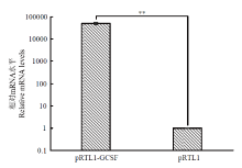
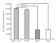
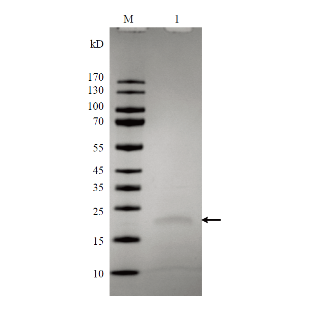
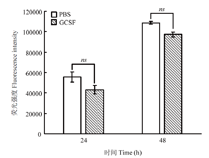
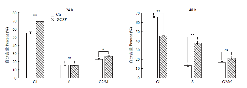
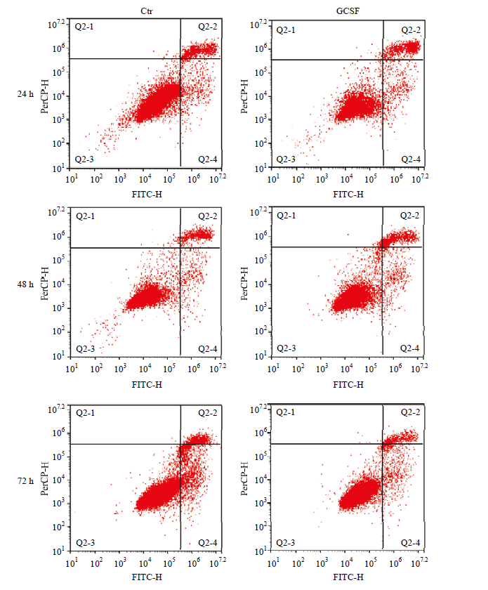
李闰婷, 陈龙欣, 张丽萌, 何海迎, 王泳, 杨若晨, 段春辉, 刘月琴, 王玉琴, 张英杰. 绵羊粒细胞集落刺激因子原核表达与提纯及用于颗粒细胞培养的效果. 生物工程学报, 2020,36(9):1817-1827. doi.org/10.13345/j.cjb.190584.
doi: 10.13345/j.cjb.190584
|
|
LI R T, CHEN L X, ZHANG L M, HE H Y, WANG Y, YANG R C, DUAN C H, LIU Y Q, WANG Y Q, ZHANG Y J. Prokaryotic expression and purification of sheep granulocyte colony stimulating factor and its effect on granulosa cell. Chinese Journal of Biotechnology, 2020,36(9):1817-1827. doi.org/10.13345/j.cjb.190584. (in Chinese)
doi: 10.13345/j.cjb.190584
|
| [25] |
BAI Y, ANN D K, SHEN W C. Recombinant granulocyte colony- stimulating factor-transferrin fusion protein as an oral myelopoietic agent. Proceedings of the National Academy of Sciences of the United States of America, 2005,102(20):7292-7296.
|
| [26] |
CUPP J S, MILLER M A, MONTGOMERY K D, NIELSEN T O, O'Connell J X, HUNTSMAN D, van de R M, GILKS C B, WEST R B. Translocation and expression of CSF1 in pigmented villonodular synovitis, tenosynovial giant cell tumor, rheumatoid arthritis and other reactive synovitides. The American Journal of Surgical Pathology, 2007,31(6):970-976.
doi: 10.1097/PAS.0b013e31802b86f8
|
| [27] |
ZHAI S Z, GUO H D, LI S Q, ZHAO X S, WANG Y, XU L P, LIU K Y, HUANG X J, CHANG Y J. Effects of granulocyte colony- stimulating factor on proliferation and apoptosis of B cells in bone marrow of healthy donors. Transplantation Proceedings, 2020,52(1):345-352.
|
| [28] |
MICKIENE G, DALGEDIENE I, DAPKUNAS Z, ZVIRBLIS G, PESLIAKAS H, KAUPINIS A, VALIUS M, MISTINIENE E, PLECKAITYTE M. Construction, purification, and characterization of a Homodimeric Granulocyte colony-stimulating factor. Molecular Biotechnology, 2017,59(9/10):374-384.
doi: 10.1007/s12033-017-0026-7
|
| [29] |
YAN J J, RYU J H, PIAO H, HWANG J H, HAN D, LEE S K, JANG J Y, LEE J, KOO T Y, YANG J. Granulocyte colony-stimulating factor attenuates renal ischemia-reperfusion injury by inducing myeloid- derived suppressor cells. Journal of the American Society of Nephrology, 2020,31(4):731-746.
doi: 10.1681/ASN.2019060601
|
| [30] |
BROCKMEIER S L, LOVING C L, EBERLE K C, HAU S J, MOU K T, KEHRLI M E JR. Administration of granulocyte-colony stimulating factor (G-CSF) to pigs results in a longer mean survival time after exposure to Streptococcus suis. Veterinary Microbiology, 231:116-119.
doi: 10.1016/j.vetmic.2019.03.010
|
| [31] |
ARMENISE A, TREROTOLI P, CIRONE F, DE NITTO A, DE SARIO C, BERTAZZOLO W, PRATELLI A, DECARO N. Use of recombinant canine granulocyte-colony stimulating factor to increase leukocyte count in dogs naturally infected by canine parvovirus. Veterinary Microbiology, 2019,231:177-182.
doi: 10.1016/j.vetmic.2019.03.015
|
| [32] |
FRASER J K, GUERRA J J, NGUYEN C Y, INDES J E, GASSON J C, NIMER S D. Characterization of a cell-type-restricted negative regulatory activity of the human granulocyte-macrophage colony- stimulating factor gene. Molecular and Cellular Biology, 1994,14(3):2213-2221.
|
| [33] |
PYKHTINA M B, ROMANOV V P, MIROSHNICHENKO S M, BEKLEMISHEV A B. Construction of a Pichia pastoris strain efficiently producing recombinant human granulocyte-colony stimulating factor (rhG-CSF) and study of its biological activity on bone marrow cells. Molecular Biology Reports, 2020,47(1):607-620.
doi: 10.1007/s11033-019-05169-9
|
| [34] |
付瑶. 人粒细胞集落刺激因子的基因合成、原核表达与活性研究[D]. 长春: 吉林大学, 2011.
|
|
FU Y. Gene synthesis, prokaryotic expression and activity of human granulocyte colony stimulating factor[D]. Changchun: Jilin University, 2011. (in Chinese)
|
| [35] |
WU Y F, GU M H, YANG S H, WANG T F. Lower platelet count with increased density of platelet antigens in granulocyte colony- stimulating factor mobilized peripheral blood stem cell donors. Journal of the Formosan Medical Association, 2020,119(1 Pt 2):204-210.
doi: 10.1016/j.jfma.2019.04.003
|
| [36] |
STEPHENS J M, BENSINK M, BOWERS C, HOLLENBEAK C S. Risks and consequences of travel burden on prophylactic granulocyte colony-stimulating factor administration and incidence of febrile neutropenia in an aged medicare population. Current Medical Research and Opinion, 2019,35(2):229-240.
doi: 10.1080/03007995.2018.1465906
|
| [37] |
MODI J, MENZIE-SUDERAM J, XU H, TRUJILLO P, MEDLEY K, MARSHALL M L, TAO R, PRENTICE H, WU J Y. Mode of action of granulocyte-colony stimulating factor (G-CSF) as a novel therapy for stroke in a mouse model. Journal of Biomedical Science, 2020,27(1):19.
doi: 10.1186/s12929-019-0597-7
|
| [38] |
PUTZ E J, EDER J M, REINHARDT T A, SACCO R E, CASAS E, LIPPOLIS J D. Differential phenotype of immune cells in blood and milk following pegylated granulocyte colony-stimulating factor therapy during a chronic Staphylococcus aureus infection in lactating Holsteins. Journal of Dairy Science, 2019,102(10):9268-9284.
doi: 10.3168/jds.2019-16448
|
| [39] |
DENIZ Y K, BURCU A, AKKIZ Ş Y, Özdemir H H, SIVIŞ Ö Z, HüSEYIN O, FERDA Ö. Congenital neutropenia patient with hypomorphic biallelic CSF3R mutation responding to GCSF. Journal of Pediatric Hematology/Oncology, 2019,41(3):190-192.
|
| [40] |
关雪莲, 侯丽淳, 杨慧, 胡婧, 王辰, 王秀萍, 毕胜, 许晓燕, 王昆祥, 和梅. G-CSF对大鼠脑出血周边组织bcl-2、caspase-3表达的影响. 中风与神经疾病杂志, 2015,32(6):511-513.
|
|
GUAN X L, HOU L C, YANG H, HU J, WANG C, WANG X P, BI S, XU X Y, WANG K X, HE M. Effects of G-CSF on expression of bcl-2 and caspase-3 around hematoma on acute intracerebral hemorrhage (ICH) in rats. Journal of Apoplexy and Nervous Diseases, 2015,32(6):511-513. (in Chinese)
|
| [41] |
姚一龙, 李隐侠, 安外尔·热合曼, 张俊, 孟春花, 王慧利, 钱勇, 曹少先. 湖羊Sp1基因CDS区克隆及其对颗粒细胞增殖和凋亡的影响. 畜牧兽医学报, 2017,48(11):2098-2106.
|
|
YAO Y L, LI Y X, REHEMAN·A, ZHANG J, MENG C H, WANG H L, QIAN Y, CAO S X. Cloning of Sp1 Gene CDS region of Hu sheep and its effect on proliferation and apoptosis of granulosa cell. Chinese Journal of Animal and Veterinary Sciences, 2017,48(11):2098-2106. (in Chinese)
|


 ),陈龙欣2,张丽萌1,2,何海迎1,王泳1,杨若晨1,段春辉1,刘月琴1,王玉琴3,张英杰1(