Scientia Agricultura Sinica ›› 2026, Vol. 59 ›› Issue (15): 3452-3464.doi: 10.3864/j.issn.0578-1752.2026.15.016

• ANIMAL SCIENCE·VETERINARY SCIENCE • Previous Articles    

Establishment of an Infection Model in Pigs for Chinese Clinical Strains of Lawsonia intracellularis

XIE Rui1(), GU ZhengQuan1, LIU SiWen1, LUO YaJuan1, CUI YuCong1, HUA Lin1, SONG WenBo2, HUANG Chao2, TANG XiBiao2, XU GaoYuan2, CHEN HuanChun1, WU Bin1(), PENG Zhong1()   

  1. 1 College of Veterinary Medicine Huazhong Agricultural University/Hubei Hongshan Laboratory/The Cooperative Innovation Center for Sustainable Pig Production, Wuhan 430070
    2 Wuhan Keqian Biology Co., Ltd., Wuhan 430073
  • Received:2026-01-29 Accepted:2026-06-04 Online:2026-08-01 Published:2026-08-03
  • Contact: WU Bin, PENG Zhong

Abstract:

【Objective】This study aimed to establish a stable porcine proliferative enteropathy (PPE) model using Chinese isolates of Lawsonia intracellularis (LI) and clinical infected intestinal tissue homogenates from different sources, construct a systematic evaluation index system covering fecal bacterial shedding, fecal score, average daily gain, feed conversion ratio, serum antibody level and histopathological changes, and identify the optimal challenge dose and preferred strain, so as to provide the standardized model and data support for the research on LI pathogenic mechanism, vaccine efficacy evaluation and prevention and control strategies.【Method】Twenty-eight-day-old weaned piglets were selected and divided into a negative control group and multiple infection groups. In Experiment 1, the pathogenicity of four LI tissue homogenates (JX703, GX301, HN312, and LI-HuB23) was compared at the dose of 1×10⁹ LI per piglet. In Experiment 2, JX703 tissue homogenate with the strongest comprehensive pathogenicity and the in vitro cultured LI-HuB23 isolate were selected to orally inoculate 28-day-old piglets at gradient doses (1010 LI per pig, 108 LI per pig, and 106 LI per pig). The piglets were monitored for 28 days after inoculation, during which fecal bacterial shedding, fecal score, average daily gain, feed conversion ratio, serum antibody level and histopathological changes were observed and recorded.【Result】In Experiment 1, typical clinical symptoms and pathological manifestations of PPE were successfully reproduced in all infection groups inoculated with tissue homogenates of different LI strains. LI was detectable in feces from day 4 post-inoculation, and bacterial shedding persisted until day 28 post-inoculation. Diarrhea occurred in the infection groups from day 4 post-inoculation, with the most severe symptoms observed from day 19 to 21 post-inoculation. All infection groups showed lower average daily gain and higher feed conversion ratio compared with the negative control group. Serum antibody monitoring showed that seroconversion occurred from day 14 after the establishment of infection and lasted until day 28 post-inoculation. Pathological examination revealed thickened intestinal mucosal folds in the infection groups; hematoxylin-eosin (HE) staining showed intestinal crypt hyperplasia and decreased goblet cells, and LI was detected in the cytoplasm of intestinal crypt epithelial cells by immunohistochemical staining (IHC). Overall, JX703 tissue homogenate induced the most prominent symptoms with the strongest virulence. In Experiment 2, the results of gradient dose test showed that inoculation with LI-HuB23 in 106 LI per piglet was sufficient to establish effective infection, accompanied by fecal bacterial shedding, diarrhea, and corresponding pathological changes. The severity of symptoms increased with the elevation of infection dose, and the 1010 LI per pig group exhibited the most typical patterns in terms of peak fecal scores, pathological scores, and reductions in average daily gain.【Conclusion】A PPE infection model was successfully established using Chinese LI isolates and field-derived infected tissue homogenates in this study. The selected evaluation indexes had an important reference value for related research, including fecal bacterial shedding, fecal score, average daily gain, feed conversion ratio, serum antibody level and histopathological changes. The infection model established with an inoculation dose of 1010 LI per pig presented more typical clinical and pathological characteristics of PPE.

Key words: porcine proliferative enteropathy, Lawsonia intracellularis, swine, infection model, challenge dose

Fig. 1

Monitoring of indicators in pigs after infection with different tissue homogenates of LI A: Fecal LI content at different time points post-infection; B: Fecal consistency scores of pigs in each group at different time points post-infection; C: Average daily gain (ADG) of pigs in each group across different infection stages; D: Representative fecal morphology of diseased pigs: (a) Watery feces, (b) Soft feces. Note: Negative control represents pigs orally administered with sterile SPG; JX703, GX301, HN312, and LI-HuB23 represent pigs challenged with different LI tissue homogenates, respectively. Data are shown as mean±SD. *P<0.05, ** P<0.01, *** P<0.001, **** P<0.0001 versus the negative control group at the matched time point"

Table 1

Feed conversion ratio of experimental pigs in each group (kg·kg-1)"

时间 Time 阴性对照 Negative control JX703 GX301 HN312 LI-HuB23
0-28d 2.66±0.39 3.65±1.14 3.47±0.56 3.91±1.30 3.13±0.47

Fig. 2

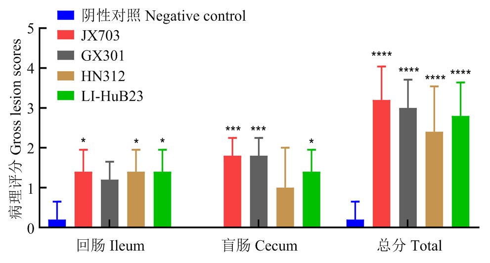
Pathological scores of intestinal tissues in experimental pigs Negative control represents pigs orally administered with sterile SPG; JX703, GX301, HN312, and LI-HuB23 represent pigs challenged with different LI tissue homogenates, respectively. *P<0.05, ** P<0.01, *** P<0.001, **** P<0.0001 versus the negative control group at the matched time point"

Fig. 3

Gross and histopathological changes of intestinal tissues in experimental pigs Yellow arrows in H.E staining indicate crypt hyperplasia and goblet cell reduction; red arrows in IHC staining indicate LI colonization. Scale bar =100 μm. Negative Control represents pigs orally administered with sterile SPG; JX703, GX301, HN312, and LI-HuB23 represent pigs challenged with different LI tissue homogenates, respectively"

Fig. 4

Preparation of LI antibody detection plate Nuclei were stained with DAPI (blue), and LI was labeled with specific green fluorescence"

Fig. 5

Detection results of serum anti-LI antibodies in experimental pigs Nuclei were stained with DAPI (blue), and green fluorescence represents LI-specific fluorescent signal; +, positive for serum LI antibody; -, negative for serum LI antibody. Negative control represents pigs orally administered with sterile SPG; JX703, GX301, HN312, and LI-HuB23 represent pigs challenged with different LI tissue homogenates, respectively"

Fig. 6

Monitoring of indicators in pigs after infection with different doses of the LI-HuB23 isolate A: Fecal LI content at different time points post-infection; B: Fecal consistency scores of pigs in each group at different time points post-infection; C: Average daily gain (ADG) of pigs in each group across different infection stages; D: Representative fecal morphology of diseased pigs: (a) Soft feces, (b) Watery feces. Note: Negative Control represents pigs orally administered with sterile SPG; LI-HuB23-1010 LI, LI-HuB23-108 LI, and LI-HuB23-106 LI represent pigs orally challenged with LI-HuB23 isolate at the dose of 1×10¹⁰, 1×10⁸, and 1×10⁶ LI per pig, respectively; JX703-1010 LI, JX703-108 LI, and JX703-106 LI represent pigs orally challenged with JX703 tissue homogenate at the dose of 1×10¹⁰, 1×10⁸, and 1×10⁶ LI per pig, respectively. Data are shown as mean±SD. **** P<0.0001 versus the negative control group at the matched time point"

Table 2

Feed conversion ratio of pigs in each group"

时间
Time
阴性对照
Negative control
LI-HuB23
1010 LI
LI-HuB23
108 LI
LI-HuB23
106 LI
JX703
1010 LI
JX703
108 LI
JX703
106 LI
0-28d 3.09±0.57 4.03±1.02 3.69±0.43 3.64±1.29 4.44±1.09 4.58±2.83 3.19±0.55

Fig. 7

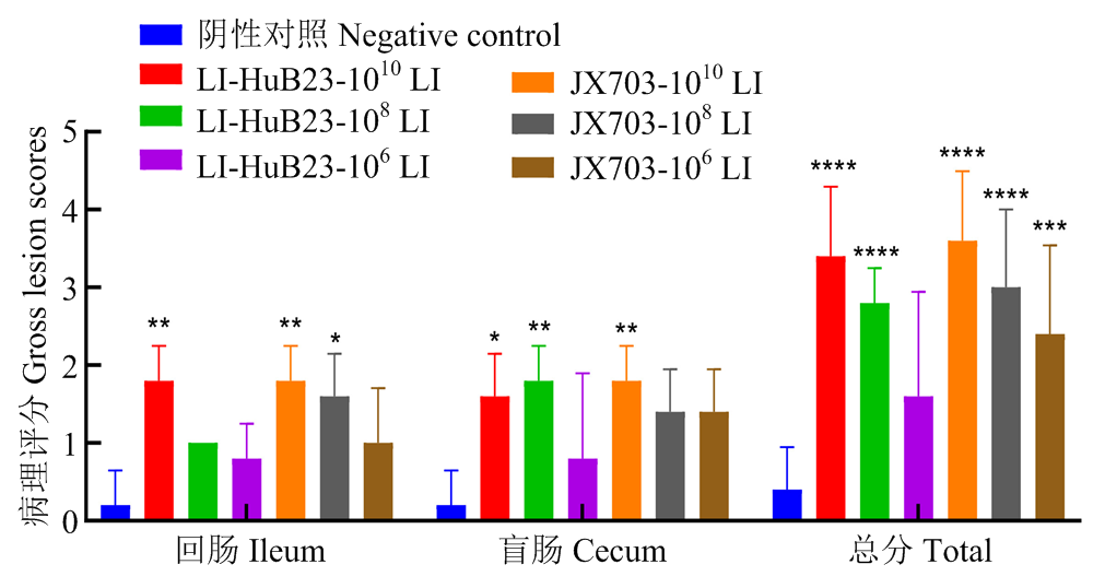
Pathological scores of intestinal tissues in experimental pigs Negative control represents pigs orally administered with sterile SPG; LI-HuB23-1010 LI, LI-HuB23-108 LI, and LI-HuB23-106 LI represent pigs orally challenged with LI-HuB23 isolate at the dose of 1×10¹⁰, 1×10⁸, and 1×10⁶ LI per pig, respectively; JX703-1010 LI, JX703-108 LI, and JX703-106 LI represent pigs orally challenged with JX703 tissue homogenate at the dose of 1×10¹⁰, 1×10⁸, and 1×10⁶ LI per pig, respectively. *P<0.05, ** P<0.01, *** P<0.001, **** P<0.0001 versus the negative control group at the matched time point"

Fig. 8

Gross and histopathological changes of intestinal tissues in experimental pigs Yellow arrows in H.E staining indicate crypt hyperplasia and goblet cell reduction; red arrows in IHC staining indicate LI colonization. Scale bar =100 μm. Negative Control represents pigs orally administered with sterile SPG; LI-HuB23-1010 LI, LI-HuB23-108 LI, and LI-HuB23-106 LI represent pigs orally challenged with LI-HuB23 isolate at the dose of 1×10¹⁰, 1×10⁸, and 1×10⁶ LI per pig, respectively; JX703-1010 LI, JX703-108 LI, and JX703-106 LI represent pigs orally challenged with JX703 tissue homogenate at the dose of 1×10¹⁰, 1×10⁸, and 1×10⁶ LI per pig, respectively"

Fig. 9

Detection results of serum anti-LI antibodies in experimental pigs Nuclei were stained with DAPI (blue), and green fluorescence represents LI-specific fluorescent signal; +, positive for serum LI antibody; -, negative for serum LI antibody. Negative control represents pigs orally administered with sterile SPG; LI-HuB23-1010 LI, LI-HuB23-108 LI, and LI-HuB23-106 LI represent pigs orally challenged with LI-HuB23 isolate at the dose of 1×10¹⁰, 1×10⁸, and 1×10⁶ LI per pig, respectively; JX703-1010 LI, JX703-108 LI, and JX703-106 LI represent pigs orally challenged with JX703 tissue homogenate at the dose of 1×10¹⁰, 1×10⁸, and 1×10⁶ LI per pig, respectively"

[1]
Lawson G H, McOrist S, Jasni S, Mackie R A. Intracellular bacteria of porcine proliferative enteropathy: Cultivation and maintenance in vitro[J]. Journal of Clinical Microbiology, 1993, 31(5): 1136-1142.
[2]
McOrist S, Gebhart C J, Boid R, Barns S M. Characterization of Lawsonia intracellularis gen. Nov., sp. Nov., the obligately intracellular bacterium of porcine proliferative enteropathy[J]. International Journal of Systematic Bacteriology, 1995, 45(4): 820-825.
[3]
McOrist S, Jasni S, Mackie R A, MacIntyre N, Neef N, Lawson G H. Reproduction of porcine proliferative enteropathy with pure cultures of ileal symbiont intracellularis[J]. Infection and Immunity, 1993, 61(10): 4286-4292.
[4]
Vannucci F A, Gebhart C J, McOrist S. Proliferative enteropathy[M]//Zimmerman J J, Karriker L A, Ramirez A, et al. Diseases of Swine. 11th ed. Ames: Wiley-Blackwell, 2019: 898-911.
[5]
Paradis M A, Gebhart C, Toole D, Vessie G, Winkelman N, Bauer S, Wilson J, McClure C. Subclinical ileitis: Diagnostic and performance parameters in a multi-dose mucosal homogenate challenge model[J]. Journal of Swine Health and Production, 2012, 20(3): 137-141.
[6]
Pedersen K S, Skrubel R, Stege H, Angen Ø, Ståhl M, Hjulsager C, Larsen L E, Nielsen J P. Association between average daily gain, faecal dry matter content and concentration of Lawsonia intracellularis in faeces[J]. Acta Veterinaria Scandinavica, 2012, 54(1): 58.
[7]
Johansen M, Nielsen M, Dahl J, Svensmark B, Bækbo P, Kristensen C S, Hjulsager C K, Jensen T K, Ståhl M, Larsen L E, Angen Ø. Investigation of the association of growth rate in grower-finishing pigs with the quantification of Lawsonia intracellularis and porcine circovirus type 2[J]. Preventive Veterinary Medicine, 2013, 108(1): 63-72.
[8]
McOrist S. Defining the full costs of endemic porcine proliferative enteropathy[J]. Veterinary Journal (London, England, 2005, 170(1): 8-9.
[9]
Veenhuizen M F, Elam T E, Soenksen N. Porcine proliferative enteropathy: Diagnosis and impact[J]. Compendium on Continuing Education for the Practising Veterinarian (North American Edition), 2002, 24 (2): S10-S15.
[10]
肖海君, 梅小伟. 2022—2023年全国主要养猪地区猪回肠炎感染情况调查[J]. 国外畜牧学(猪与禽), 2023, 43(6): 102-107.
Xiao H J, Mei X W. Investigation of Crohn’s disease infection in major pig-raising areas in China from 2022 to 2023[J]. Animal Science Abroad-Pigs and Poultry, 2023, 43(6): 102-107.(in Chinese)
[11]
Wang L, Wu W Q, Zhao L F, Zhu Z W, Yao X Z, Fan J, Chen H J, Song W B, Huang X, Hua L, Qian P, Chen H C, Peng Z, Wu B. Fecal PCR survey and genome analysis of Lawsonia intracellularis in China[J]. Frontiers in Veterinary Science, 2024, 11: 1324768.
[12]
彭长江, 谢锐, 朱占伟, 武文清, 杨豪, 孙心如, 宋晓容, 黄婧文, 宋文博, 华琳, 陈焕春. 猪增生性肠病疫苗研究进展[J]. 中国兽医学报, 2025, 45 (12): 2793-2801.
Peng C J, Xie R, Zhu Z W, Wu W Q, Yang H, Sun X R, Song X R, Huang J W, Song W B, Hua L, Chen H C. Research progress on vaccines for porcine proliferative enteropathy[J]. Chinese Journal of Veterinary Science, 2025, 45 (12): 2793-2801.(in Chinese)
[13]
Kroll J J, Roof M B, McOrist S. Evaluation of protective immunity in pigs following oral administration of an avirulent live vaccine of Lawsonia intracellularis[J]. American Journal of Veterinary Research, 2004, 65(5): 559-565.
[14]
Hankel J, Sander S, Muthukumarasamy U, Strowig T, Kamphues J, Jung K, Visscher C. Microbiota of vaccinated and non-vaccinated clinically inconspicuous and conspicuous piglets under natural Lawsonia intracellularis infection[J]. Frontiers in Veterinary Science, 2022, 9: 1004506.
[15]
Xie R, Luo Y J, Peng C J, Wu W Q, Cui Y C, Sun X R, Hua L, Wu B, Peng Z. Isolation, passage, and pathogenicity of a newly isolated Lawsonia intracellularis strain from Hubei, China[J]. Transboundary and Emerging Diseases, 2025, 2025: 2501719.
[16]
Xiao N, Li J N, Li M X, Zhou H, Lin H X, Fan H J. Isolation and in vitro cultivation of Lawsonia intracellularis from China[J]. Veterinary Microbiology, 2022, 270: 109457.
[17]
Percie du Sert N, Ahluwalia A, Alam S, Avey M T, Baker M, Browne W J, Clark A, Cuthill I C, Dirnagl U, Emerson M, Garner P, Holgate S T, Howells D W, Hurst V, Karp N A, Lazic S E, Lidster K, MacCallum C J, MacLeod M, Pearl E J, et al. Reporting animal research: Explanation and elaboration for the ARRIVE guidelines 2.0[J]. PLoS Biology, 2020, 18(7): e3000411.
[18]
Wattanaphansak S, Gebhart C J, Anderson J M, Singer R S. Development of a polymerase chain reaction assay for quantification of Lawsonia intracellularis[J]. Journal of Veterinary Diagnostic Investigation, 2010, 22(4): 598-602.
[19]
Wu W Q, Wang L, Xie R, Peng C J, Yang H, Zhu H C, Luo Y J, Hua L, Chen H C, Wu B, Peng Z. Development and application of a triplex real-time PCR method for the detection of Lawsonia intracellularis, Brachyspira hyodysenteriae, and Clostridium perfringens[J]. Microbiology Spectrum, 2025, 13(6): e0323724.
[20]
Guedes R M, Gebhart C J, Deen J, Winkelman N L. Validation of an immunoperoxidase monolayer assay as a serologic test for porcine proliferative enteropathy[J]. Journal of Veterinary Diagnostic Investigation, 2002, 14(6): 528-530.
[21]
Chagas S, Jensen P, Paladino E, Pascoal L M, von Berg S, Gebhart C, Vannucci F A. Immune response of pigs vaccinated against proliferative enteropathy and co-infected with Lawsonia intracellularis and Brachyspira hyodysenteriae[J]. Animals, 2025, 16(1): 114.
[22]
Joens L A, Nibbelink S, Glock R D. Induction of gross and microscopic lesions of porcine proliferative enteritis by Lawsonia intracellularis[J]. American Journal of Veterinary Research, 1997, 58(10): 1125-1131.
[23]
Guedes R M C, Gebhart C J. Comparison of intestinal mucosa homogenate and pure culture of the homologous Lawsonia intracellularis isolate in reproducing proliferative enteropathy in swine[J]. Veterinary Microbiology, 2003, 93(2): 159-166.
[24]
Lin H X, Liu X, Yuan J Z, Xiao N, Zhou H, Fan H J. Evaluation of a newly developed live attenuated vaccine candidate against Lawsonia intracellularis[J]. Vaccines, 2025, 14(1): 15.
[25]
Helm E T, Burrough E R, Gabler N K, Leite F L. Lawsonia intracellularis infection induces changes in microbial community function and composition associated with reduced pig growth and feed efficiency[J]. Animal Microbiome, 2026, 8(1): 1.
[26]
Smith S H, McOrist S. Development of persistent intestinal infection and excretion of Lawsonia intracellularis by piglets[J]. Research in Veterinary Science, 1997, 62(1): 6-10.
[27]
Guedes R M C, Gebhart C J. Onset and duration of fecal shedding, cell-mediated and humoral immune responses in pigs after challenge with a pathogenic isolate or attenuated vaccine strain of Lawsonia intracellularis[J]. Veterinary Microbiology, 2003, 91(2/3): 135-145.
[28]
Vannucci F A, Beckler D, Pusterla N, Mapes S M, Gebhart C J. Attenuation of virulence of Lawsonia intracellularis after in vitro passages and its effects on the experimental reproduction of porcine proliferative enteropathy[J]. Veterinary Microbiology, 2013, 162(1): 265-269.
[29]
Guedes R M C, Machuca M A, Quiroga M A, Pereira C E R, Resende T P, Gebhart C J. Lawsonia intracellularis in pigs: Progression of lesions and involvement of apoptosis[J]. Veterinary Pathology, 2017, 54(4): 620-628.
[30]
Wattanaphansak S, Pereira C E R, Kaenson W, Assavacheep P, Tantilertcharoen R, Resende T P, Barrera-Zarate J A, de Oliveira-Lee J S V, Klein U, Gebhart C J, Guedes R M C. Isolation and in vitro antimicrobial susceptibility of porcine Lawsonia intracellularis from Brazil and Thailand[J]. BMC Microbiology, 2019, 19(1): 27.
[31]
Vannucci F A, Wattanaphansak S, Gebhart C J. An alternative method for cultivation of Lawsonia intracellularis[J]. Journal of Clinical Microbiology, 2012, 50(3): 1070-1072.
[32]
Collins A M, Swift I, Monckton R P. Replication of Australian porcine isolates of Ileal symbiont intracellularis in tissue culture[J]. Veterinary Microbiology, 1996, 49(3/4): 249-255.
[33]
Daniel A G S, Pereira C E R, Dorella F, Pereira F L, Laub R P, Andrade M R, Barrera-Zarate J A, Gabardo M P, Otoni L V A, Macedo N R, Correia P A, Costa C M, Vasconcellos A O, Wagatsuma M M, Marostica T P, Figueiredo H C P, Guedes R M C. Synergic effect of Brachyspira hyodysenteriae and Lawsonia intracellularis coinfection: Anatomopathological and microbiome evaluation[J]. Animals, 2023, 13(16): 2611.
[34]
Meschede J, Holtrup S, Deitmer R, Mesu A P, Kraft C. Reduction of Salmonella prevalence at slaughter in Lawsonia intracellularis co-infected swine herds by Enterisol® Ileitis vaccination[J]. Heliyon, 2021, 7(4): e06714.
[35]
McORIST S, Lawson G H K. Reproduction of proliferative enteritis in gnotobiotic pigs[J]. Research in Veterinary Science, 1989, 46(1): 27-33.
[36]
Wattanaphansak S, Singer R S, Gebhart C J. In vitro antimicrobial activity against 10 North American and European Lawsonia intracellularis isolates[J]. Veterinary Microbiology, 2009, 134(3/4): 305-310.
[1] HONG RunJing, ZHOU Hong, LIN HuiXing, FAN HongJie. Establishment and Application of Sandwich ELISA Method for Detecting Lawsonia intracellularis [J]. Scientia Agricultura Sinica, 2025, 58(5): 1032-1042.
[2] HE XiaoPing, ZHANG YuanFeng, LIU XueMin, HUANG Li, WENG ChangJiang. Preparation of Monoclonal Antibody Against African Swine Fever Virus pD345L Protein and Identification of Its Epitope [J]. Scientia Agricultura Sinica, 2025, 58(16): 3345-3356.
[3] WAN YunFei, YANG YuYing, ZHANG NaiXin, XU MengMeng, YU QinHao, QIAO ChuanLing, CHEN HuaLan. Identification and Functional Analysis of Adaptive Amino Acid Mutations in the Eurasian Avian-Like H1N1 Swine Influenza Virus [J]. Scientia Agricultura Sinica, 2025, 58(10): 2035-2044.
[4] LIU ChuanXia, CHEN Xin, WANG Xiao, LI XueWen, LI TingTing, WENG ChangJiang, ZHENG Jun. Preparation and Application of Polyclonal Antibodies Against Pig CD1d Protein [J]. Scientia Agricultura Sinica, 2024, 57(8): 1620-1628.
[5] ZHAO YiRan, SHAN YanKe, LI JiaHao, HE ZhaoQun, WANG XinYi, WEN Dun, WANG MiLa, CHU Rui, ZHAO DongMing, LIU Fei. Establishment of Rapid Field Co-Detection Method of ASFV Antibody and Nucleic Acid Based on Quantum Dot Microspheres and RPA Technology [J]. Scientia Agricultura Sinica, 2024, 57(24): 4990-5002.
[6] ZHAO WenShuo, ZHANG JinLong, YAO ZhaoRan, SONG YuQi, LÜ Shun, LIU YingXue, YUAN CongCong, SUN YuHang. Effects of Aflatoxin B1 on Influenza Virus Replication, Organ Damages and Intestinal Microbiota Disorder of Swine [J]. Scientia Agricultura Sinica, 2024, 57(20): 4145-4160.
[7] FENG ChunYing, ZHANG ZhaoXia, LIU YunFei, HUANG Li, WENG ChangJiang. Preparation of Monoclonal Antibody Against African Swine Fever Virus p54 Protein and Identification of Its Epitope [J]. Scientia Agricultura Sinica, 2024, 57(19): 3936-3944.
[8] ZHANG CongYue, ZHOU Hong, LIN HuiXing, FAN HongJie. Development and Application of Indirect ELISA Kits for Antibody Detection of Porcine Proliferative Enteropathy [J]. Scientia Agricultura Sinica, 2024, 57(16): 3283-3293.
[9] FAN Shuai, ZHONG Han, YANG ZhongYuan, HE WenRui, WAN Bo, WEI ZhanYong, HAN ShiChong, ZHANG GaiPing. African Swine Fever Virus MGF110-5L-6L Induces Host Cell Translation Arrest and Stress Granule Formation by Activating the PERK/PKR-eIF2α Pathway [J]. Scientia Agricultura Sinica, 2023, 56(7): 1401-1416.
[10] ZHANG NaiXin, XU ChengZhi, YANG YuYing, ZHANG YaPing, WAN YunFei, QIAO ChuanLing, CHEN HuaLan. Identification of Key Amino Acids in the Antigenic Variation of Eurasian Avian-Like H1N1 Swine Influenza Viruses [J]. Scientia Agricultura Sinica, 2023, 56(14): 2828-2836.
[11] HUANG Cheng, YANG LongFeng, SUN Peng, CHENG HuiMin, YANG ZhiYuan, LIN Jian, ZHU HongWei, LIU LiXin, SUN HouMin, LI JiaFeng, ZHAO JiCheng, DUAN HuiJuan, PAN Jie, LIU YueHuan. Establishment of a Canine Experimental Infection Model with a H3N2 Subtype Canine Influenza Virus [J]. Scientia Agricultura Sinica, 2023, 56(13): 2620-2628.
[12] WANG Tao, LUO Rui, SUN Yuan, QIU HuaJi. Development Strategies and Application Prospects of African Swine Fever Vaccines: Feasibility and Probability [J]. Scientia Agricultura Sinica, 2023, 56(11): 2212-2222.
[13] WANG YiDan,YANG FaLong,CHEN DiShi,XIANG Hua,REN YuPeng. One-Step Multiple TaqMan Real-time RT-PCR for Simultaneous Detection of Swine Diarrhea Viruses [J]. Scientia Agricultura Sinica, 2023, 56(1): 179-192.
[14] ZHAI XiaoHu, LI LingXu, CHEN XiaoZhu, JIANG HuaiDe, HE WeiHua, YAO DaWei. Quantitative Detection Technology of Porcine-Derived Materials in Meat by Real-time PCR [J]. Scientia Agricultura Sinica, 2023, 56(1): 156-164.
[15] YANG ShiMan, XU ChengZhi, XU BangFeng, WU YunPu, JIA YunHui, QIAO ChuanLing, CHEN HuaLan. Amino Acid of 225 in the HA Protein Affects the Pathogenicities of H1N1 Subtype Swine Influenza Viruses [J]. Scientia Agricultura Sinica, 2022, 55(4): 816-824.
Viewed
Full text


Abstract

Cited

  Shared   
  Discussed   
No Suggested Reading articles found!