Scientia Agricultura Sinica ›› 2026, Vol. 59 ›› Issue (10): 2276-2287.doi: 10.3864/j.issn.0578-1752.2026.10.015

• ANIMAL SCIENCE·VETERINARY SCIENCE • Previous Articles     Next Articles

Establishment of Fluorescence Quantitative PCR Detection Method for Mycoplasma Contamination in Cells and Virus Live Vaccines

LIU Dan1,2(), GAO JianShuai1(), QIAN JiaHao1, ZHANG BoYuan1, LI HuiTong1,2, DING JiaBo1, XIONG Tao2(), SHEN QingChun1()   

  1. 1 Key Laboratory of Animal Biosafety Risk Prevention and Control (North)/China Institute of Animal Sciences, Chinese Academy of Agricultural Sciences, Beijing 100193
    2 College of Animal Science and Technology, Yangtze University, Jingzhou 434025, Hubei
  • Received:2025-03-09 Accepted:2026-04-07 Online:2026-05-16 Published:2026-05-20
  • Contact: XIONG Tao, SHEN QingChun

Abstract:

【Background】Mycoplasma contamination poses a persistent challenge in biological research and biopharmaceutical production. Conventional detection methods, such as classical culture assays, are time-consuming and lack sensitivity, while the standard PCR is prone to inhibitor interference and incapable of quantification, failing to meet the demand for rapid and precise quality control in vaccine production or laboratory settings.【Objective】This study aimed to establish a universal, highly specific, and sensitive quantitative real-time PCR (qPCR) method for efficient screening of mycoplasma contamination in cell cultures, live viral vaccines, and biological raw materials.【Method】The SILVA_138.1_SSURef database, encompassing 16S/18S rRNA sequences of bacteria, archaea, and fungi, was utilized to extract 181 non-redundant 16S rRNA sequences from classified mycoplasma species. The hypervariable V6-V8 region was identified as the optimal target via multiple sequence alignment (MEGA 11.0). Three primers (forward primers MF1: 5′-GCAAARCTATRGARAYATAGYVGAG-3′; MF2: 5′-GCAAAGGCT TAGAAATAAGTTCGGAG-3; reverse primer MR: 5′-CCARCTCYCATRGTKTGACGG-3′) and a dual-quenched TaqMan probe (5′-FAM-ACAGRTGGTGCATGGYTGTCGTCAGCTC-BHQ1-3′) were designed using Primer Premier 5.0, with primer-probe ratios and annealing temperatures optimized to establish the qPCR assay. Validation included: (1) primer-probe specificity testing against 11 Mycoplasma/Acholeplasma species (e.g., Mycoplasma anatis, Mycoplasma bovis, Ureaplasma urealyticum); (2) sensitivity assessment via 10-fold serial dilutions (1.0×10⁸-1.0×101 copies/μL) of Mycoplasma synoviae BHQ03, Mycoplasma gallisepticum, and Mycoplasma hyopneumoniae, with standard curves generated; (3) specificity evaluation against four common bacteria (Salmonella, Clostridium perfringens, Escherichia coli, Brucella) and eight animal cell lines (Marc145, Vero, CEF, etc.); (4) repeatability analysis (intra- and inter-assay variability) using M. synoviae BHQ03 dilutions (1.0×10⁸-1.0×101 copies/μL); (5) parallel testing of 24 live viral vaccine batches (poultry, swine, and canine), 20 cell culture samples (8 types), and 8 viral seed stocks via qPCR, conventional PCR, and classical culture methods.【Result】The optimized qPCR protocol employed a two-step amplification program (56 ℃ annealing temperature). Specificity testing confirmed positive detection of all 11 Mycoplasma/ Acholeplasma strains and no cross-reactivity with non-target bacteria or cell lines. Repeatability tests showed coefficient of variation (CV) values <2% for Ct values across replicates. Sensitivity assays demonstrated limits of detection (LOD) of 1-2 copies/μL for M. synoviae, M. gallisepticum, and M. hyopneumoniae. Comparative analysis of clinical samples revealed high concordance between qPCR, conventional PCR, and culture methods, with qPCR exhibiting superior sensitivity.【Conclusion】The universal qPCR method developed in this study provided an accurate, reliable, and rapid detection tool for potential mycoplasma contamination in cell cultures and live viral vaccines.

Key words: mycoplasma, fluorescence quantitative PCR, biological products

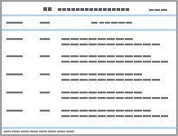
Fig. 1

16S rRNA gene sequence alignment results of mycoplasma and steroids as well as primer and probe binding regions"

Table 1

Sequences of primers and probes for fluorescent quantitative PCR"

引物及探针
Primer and probe
序列
Sequence(5′-3′)
MF1 GCAAARCTATRGARAYATAGYVGAG
MF2 GCAAAGGCTTAGAAATAAGTTCGGAG
MR CCARCTCYCATRGTKTGACGG
MyT4 FAM-ACAGRTGGTGCATGGYTGTCGTCAGCTC
-BHQ1

Table 2

Sequences of primers and probes for digital PCR"

支原体 Mycoplasma 引物及探针 Primer and probe 序列 Sequence(5′-3′)
猪肺炎支原体
M.hyopneumoniae
Mhp-F CCAGAACCAAATTCCTTCGCTG
Mhp-R ACTGGCTGAACTTCATCTGGGCTA
Mhp-P FAM-AGCAGATCTTAGTCAAAGTGCCCGTG-BHQ
鸡毒支原体
M.gallisepticum
MG-F TTGGGTTTAGGGATTGGGATT
MG-R CCAAGGGATTCAACCATC
MG-P FAM-TGATGATCCAAGAACGTGAAGAACACC-BHQ1
鸡滑液囊支原体
M.synoviae
MSD-F TGTGTTTGCGGGCTTGTATT
MSD-R TGCATTAGTTCCTGCATTAGTTCAA
MSD-P FAM-AGAAGCCGAAGCTGGAGTAGCAGT-HBQ1

Fig. 2

Results of primers and probes for real-time fluorescent quantitative PCR"

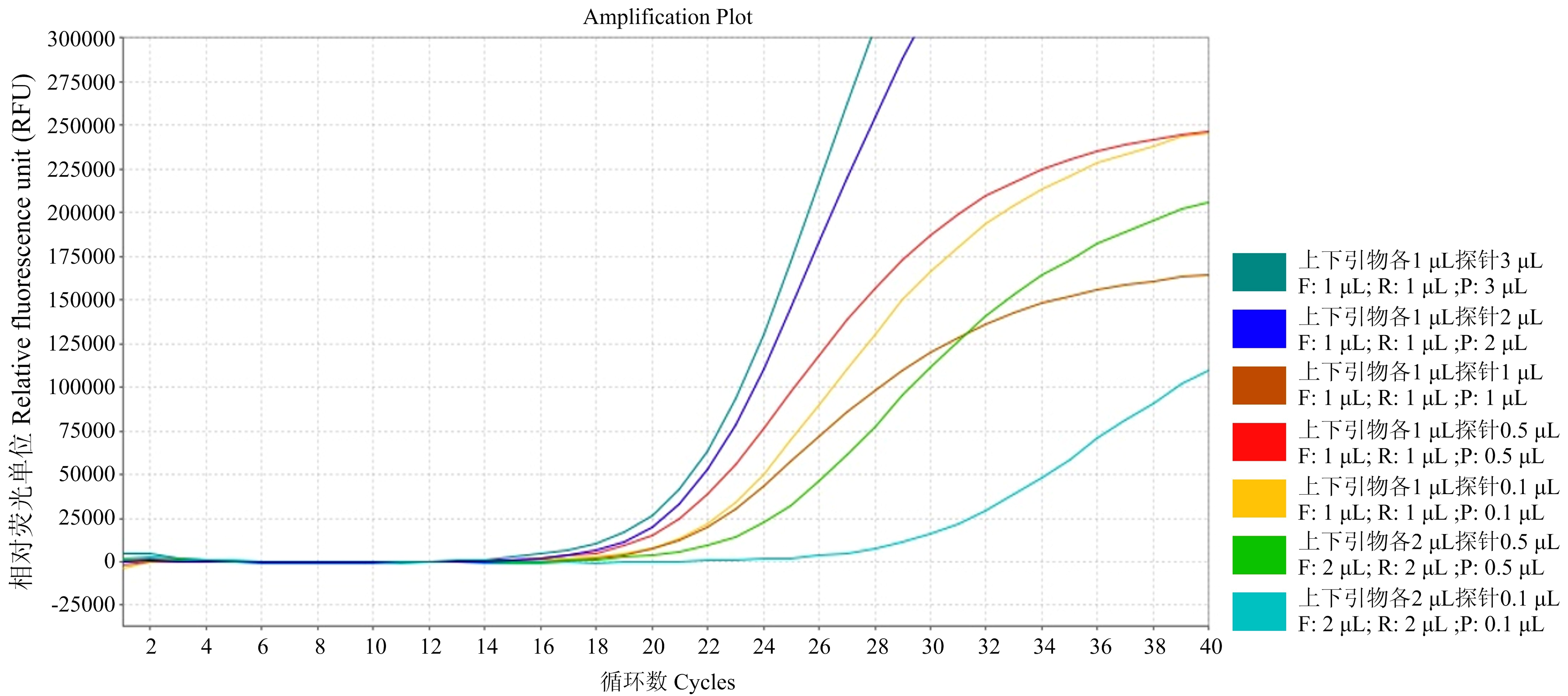
Fig. 3

Testing results of different proportions of primer and probe concentrations in Real-time fluorescent quantitative PCR method"

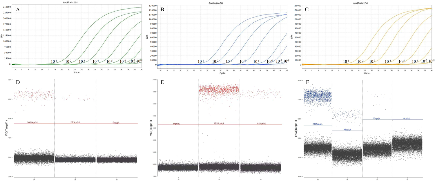
Fig. 4

qPCR sensitivity test results for mycoplasma A, B, C: Fluorescence quantitative PCR sensitivity curves for Mycoplasma synoviae strain BHQ03, Mycoplasma gallisepticum, and Mycoplasma hyopneumoniae; D, E, F: Digital PCR result graphs for Mycoplasma synoviae strain BHQ03, Mycoplasma gallisepticum, and Mycoplasma hyopneumoniae strains"

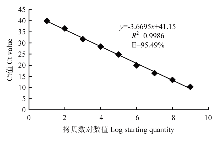
Fig. 5

Standard curve of real-time fluorescent quantitative PCR"

Fig. 6

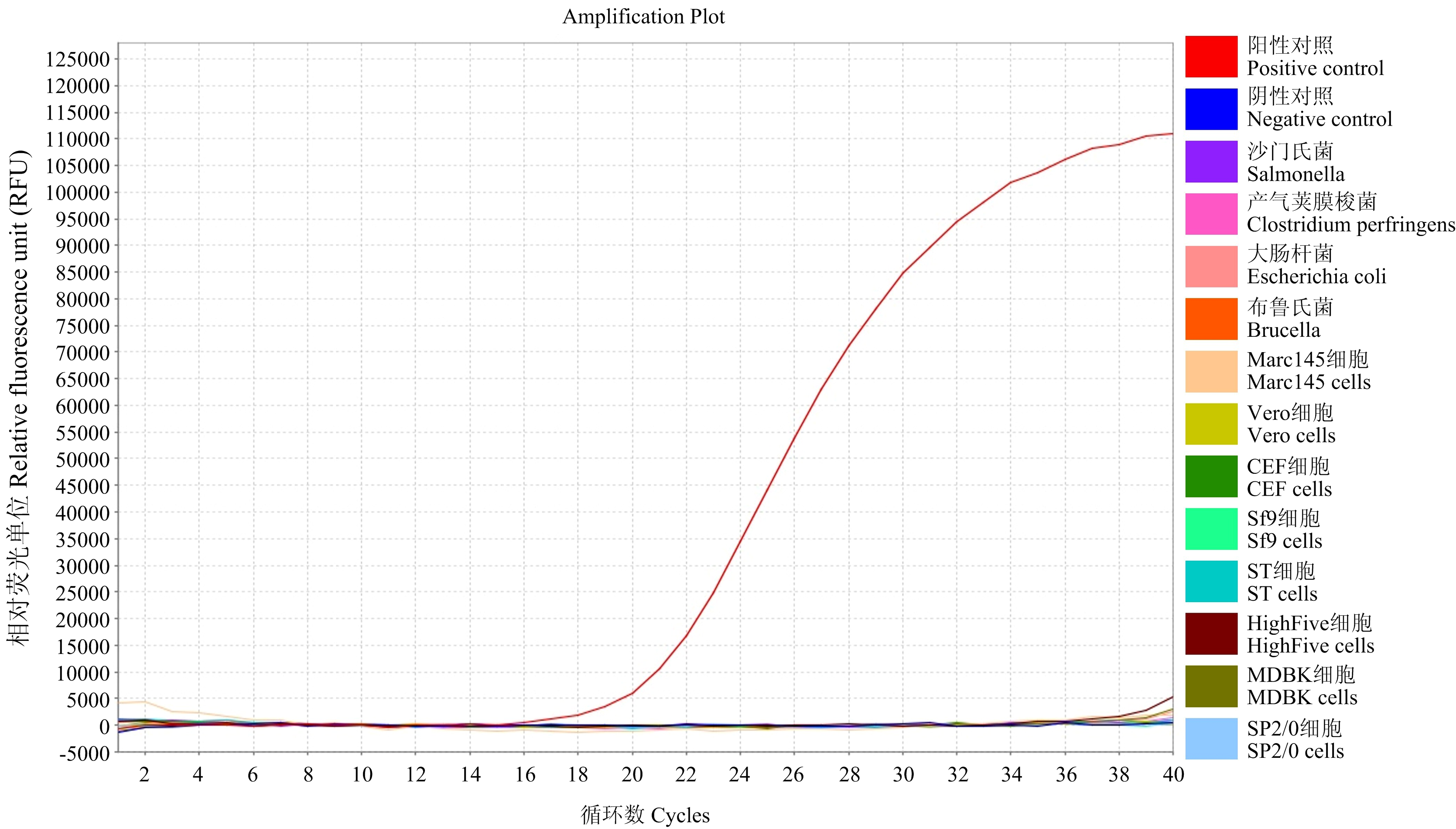
Specificity test of real-time fluorescent quantitative PCR"

Table 3

Repeatability test of real-time fluorescent quantitative PCR"

支原体
Mycoplasma
稀释倍数
Dilution (copies/μL)
Ct值1
Ct value 1
Ct值2
Ct value 2
Ct值3
Ct value 3
Ct值
Ct value (mean ± SD)
变异系数
CV (%)
鸡滑液囊支原体BHQ03株M.synoviaestrain BHQ03 1×10⁸ 10.63 10.267 10.332 10.41±0.16 1.52
1×10⁷ 13.467 13.429 13.442 13.45±0.02 0.12
1×10⁶ 16.471 16.462 16.455 16.46±0.01 0.04
1×10⁵ 19.969 19.968 19.939 19.96±0.01 0.07
1×10⁴ 22.816 22.827 22.884 22.84±0.03 0.13
1×10³ 25.373 25.295 25.522 25.40±0.09 0.37
1×10² 28.728 28.751 28.689 28.72±0.03 0.09
1×101 31.645 32.376 32.628 32.55±0.12 1.29

Table 4

The results of the veterinary live vaccines Mycoplasma detection with the PCR assay and traditional culture method"

类别
Category
生物制品名称
Biological products
原材料
Materials
阳性结果数 Number of positive samples
qPCR PCR 培养法 Cultivationa
细胞
Cells
健康细胞8株(种)
8 strains(8 species) of healthy cells
/ 0/8 0/8 0/8
待检细胞20株(8种)
20 strains(8 species) of cell samples
/ 5/20 5/20 4/20(5/20)
动物用病毒活疫苗
Live vaccines for animals
鸡用病毒活疫苗8批(品种)
8 batches (species) of live vaccines for chickens
鸡胚、CEF等
Chicken embryos,CEF,etc.
2/8 2/8 0/8
鸭用病毒活疫苗4批(品种)
4 batches (species) of live vaccines for ducks
鸭胚
Duck embryos
0/4 0/4 0/4
猪用病毒活疫苗8批(品种)
8 batches (species) of live vaccines for swine
Vero,PK15,Marc145,ST,etc. 0/8 0/8 0/8
犬用病毒活疫苗4批(品种)
4 batches (species) of live vaccines for dogs
Vero,MDCK,PK15,etc. 0/4 0/4 0/4
毒种
Virus seed
牛病毒性腹泻病毒2株(1种)
2 strains(1 species)of Bovine Viral Diarrhea Virus(BVDV)
MDBK,MDB-CX,etc. 1/2 1/2 1/2
猪细小病毒2株(2种)
2 strains(2 species)of Porcine Parvovirus(PPV)
PK15,ST,etc. 2/2 2/2 2/2
腺病毒2株(1种)
2 strains(1 species)of Adenovirus (AAV)
鸡胚、CEF等
Chicken embryos,CEF,etc.
1/2 1/2 1/2
猫杯状病毒2株(2种)
2 strains(2 species)of Feline Caliciviru(FCV)
F81,CRFK,etc. 2/2 2/2 2/2
[1]
DOUGALL A M, BRINKLEY T R, CLARKE B D. Imported biologicals: Unforeseen biosecurity risks in the laboratory. Microbiology Australia, 2020, 41(3): 132-135.

doi: 10.1071/MA20035
[2]
吴华伟, 刘丹, 王嘉, 陈晓春, 黄小洁, 孔冬妮, 苏佳, 侯力丹, 薛麒, 邓永, 翟天舒, 赵炜, 白洪旭, 薛青红. 我国禽用生物制品现状及禽用病毒类生物制品质量情况分析. 中国兽药杂志, 2024, 58(3): 30-37.
WU H W, LIU D, WANG J, CHEN X C, HUANG X J, KONG D N, SU J, HOU L D, XUE Q, DENG Y, ZHAI T S, ZHAO W, BAI H X, XUE Q H. The present situation of avain biologics and analysis on quality of avain viral biologics in China. Chinese Journal of Veterinary Drug, 2024, 58(3): 30-37. (in Chinese)
[3]
英聪, 刘灿, 王海光, 宁宜宝, 毕丁仁. 支原体污染的检测及鉴定方法研究进展. 中国家禽, 2012, 34(23): 46-49.
YING C, LIU C, WANG H G, NING Y B, BI D R. Research progress on detection and identification methods of Mycoplasma pollution. China Poultry, 2012, 34(23): 46-49. (in Chinese)
[4]
马欣, 刘博, 魏津, 王甲, 赵浩然, 刘燕, 姚文生, 罗玉峰. 国内外兽用生物制品支原体检验方法的比较分析. 中国兽药杂志, 2023, 57(5): 13-20.
MA X, LIU B, WEI J, WANG J, ZHAO H R, LIU Y, YAO W S, LUO Y F. Analysis on Mycoplasma test of veterinary biologics among Chinese veterinary pharmacopoeia, U.S. code of federal regulations and European pharmacopoeia. Chinese Journal of Veterinary Drug, 2023, 57(5): 13-20. (in Chinese)
[5]
刘轶秋, 徐磊, 张瑞婷. 兽用生物制品支原体检验项近5年抽检情况分析. 中国兽药杂志, 2012, 46(9): 45-47.
LIU Y Q, XU L, ZHANG R T. Analysis on Mycoplasma inspection of veterinary biological products sampling in recent five years. Chinese Journal of Veterinary Drug, 2012, 46(9): 45-47. (in Chinese)
[6]
国家药典委员会. 中华人民共和国药典-四部: 2015年版. 北京: 中国医药科技出版社, 2015.
Chinese Pharmacopoeia Commission. People’s Republic of China (PRC) pharmacopoeia-part IV: 2015 edition. Beijing: China Medical Science Press, 2015. (in Chinese)
[7]
中国兽药典委员会. 中华人民共和国兽药典三部 2020年版. 北京: 中国农业出版社, 2021.
Chinese Veterinary Pharmacopoeia Commission. Veterinary Pharmacopoeia of the People's Republic of China (Part III) 2020 Edition. Beijing: China Agriculture Press, 2021. (in Chinese)
[8]
Center for Veterinary Biologics (CVB). U.S. regulations and technical standards for veterinary biological products. 2019.
[9]
European Pharmacopoeia Commission. European pharmacopoeia Seventh Edition Supplement 7.1. 2010.
[10]
European Medicines Agency. VICH GL34: Biologicals: Testing for the detection of Mycoplasma contamination. 2013.
[11]
张丽君, 于佳, 韦钦钦, 石岩, 魏然, 刘晓凡. 支原体检测PCR法与药典方法的对比分析. 中国生物制品学杂志, 2020, 33(12): 1436-1440, 1444.
ZHANG L J, YU J, WEI Q Q, SHI Y, WEI R, LIU X F. Comparative analysis of PCR and method in Chinese Pharmacopoeia for Mycoplasma examination. Chinese Journal of Biologicals, 2020, 33(12): 1436-1440, 1444. (in Chinese)
[12]
全国动物卫生标准化技术委员会. 兽用生物制品外源支原体检验方法: GB/T 41699-2022. 2022.
National Technical Committee on Animal Health Standardization. Testing methods for exogenous Mycoplasma in veterinary biological products: GB/T 41699-2022. 2022. (in Chinese)
[13]
ZHU H L, ZHANG H Q, XU Y, LAŠŠÁKOVÁ S, KORABEČNÁ M, NEUŽIL P. PCR past, present and future. BioTechniques, 2020, 69(4): 317-325.

doi: 10.2144/btn-2020-0057
[14]
HOPERT A, UPHOFF C C, WIRTH M, HAUSER H, DREXLER H G. Specifity and sensitivity of polymerase chain reaction (PCR) in comparison with other methods for the detection of Mycoplasma contamination in cell lines. Journal of Immunological Methods, 1993, 164(1): 91-100.

doi: 10.1016/0022-1759(93)90279-G
[15]
DREXLER H G, UPHOFF C C. Mycoplasma contamination of cell cultures: Incidence, sources, effects, detection, elimination, prevention. Cytotechnology, 2002, 39(2): 75-90.

doi: 10.1023/A:1022913015916
[16]
YOUNG L, SUNG J, STACEY G, MASTERS J R. Detection of Mycoplasma in cell cultures. Nature Protocols, 2010, 5(5): 929-934.

doi: 10.1038/nprot.2010.43
[17]
SIMIONATTO S, MARCHIORO S B, DOS SANTOS BARBOSA M, GALLI V, BRUM C B, JORGE S, DELLAGOSTIN O A. Development of ELISA using recombinant proteins for the diagnosis of Mycoplasma hyopneumoniae infection. Indian Journal of Microbiology, 2022, 62(1): 88-95.

doi: 10.1007/s12088-021-00981-z
[18]
KHURANA S K. Detection of Mycoplasma equigenitalium from equines by capture- ELISA. Advances in Animal and Veterinary Sciences, 2015, 3(1s): 33-37.
[19]
耿仁浩, 刘博, 王芳, 罗玉峰, 曲鸿飞, 范学政, 秦玉明, 丁家波, 许冠龙, 沈青春, 秦爱建. 细胞和病毒活疫苗中支原体污染的PCR检测方法建立与应用. 中国农业科学, 2022, 55(7): 1458-1468. doi: 10.3864/j.issn.0578-1752.2022.07.016.
GENG R H, LIU B, WANG F, LUO Y F, QU H F, FAN X Z, QIN Y M, DING J B, XU G L, SHEN Q C, QIN A J. Establishment and application of PCR assay for Mycoplasma contamination in cell culture and live virus vaccine. Scientia Agricultura Sinica, 2022, 55(7): 1458-1468. doi: 10.3864/j.issn.0578-1752.2022.07.016. (in Chinese)
[20]
DOBROVOLNY P L, BESS D. Optimized PCR-based detection of Mycoplasma. Journal of Visualized Experiments, 2011(52): e3057.
[21]
WAN Q Y, LI W, ZHAO M Y, WANG H X, LI Y, SHI C, Performance analysis of novel nucleic acid detection kit for Mycoplasma pneumoniae. Clinical Pediatrics, 2022, 61(4): 330-334.

doi: 10.1177/00099228221078421
[22]
KRZYSZTOŃ-RUSSJAN J, CHUDZIAK J, BEDNAREK M, ANUSZEWSKA E L. Development of new PCR assay with SYBR green I for detection of Mycoplasma, Acholeplasma, and Ureaplasma sp. in cell cultures. Diagnostics, 2021, 11(5): 876.

doi: 10.3390/diagnostics11050876
[23]
TOTTEN A H, ADAMS A J, HALAS H K, GEBO J E T, EAST A D, LAU A F. Comparison of five commercial molecular assays for Mycoplasma testing of cellular therapy products. Journal of Clinical Microbiology, 2023, 61(2): e01498-e01422.
[24]
CARRILLO-ÁVILA J A, DE LA FUENTE A, AGUILAR-QUESADA R, LIGERO G, DEL RÍO-ORTIZ J M, CATALINA P. Development and evaluation of a new qPCR assay for the detection of Mycoplasma in cell cultures. Current Issues in Molecular Biology, 2023, 45(8): 6903-6915.

doi: 10.3390/cimb45080435
[25]
赵立群, 邱艳红, 张晓飞, 刘慧, 杨静静, 张建, 张海军, 徐秀兰, 温常龙. TaqMan探针法实时荧光定量PCR检测西瓜潜隐病毒. 中国农业科学, 2021, 54(20): 4337-4347. doi: 10.3864/j.issn.0578-1752.2021.20.008.
ZHAO L Q, QIU Y H, ZHANG X F, LIU H, YANG J J, ZHANG J, ZHANG H J, XU X L, WEN C L. The detection of Citrullus lanatus cryptic virus using TaqMan-qPCR method. Scientia Agricultura Sinica, 2021, 54(20): 4337-4347. doi: 10.3864/j.issn.0578-1752.2021.20.008. (in Chinese)
[26]
周云雷, 魏飞龙, 李健, 张小华, 蒋红霞. 鸡毒支原体实时荧光定量PCR检测方法的建立. 中国农业科学, 2011, 44(11): 2371-2378. doi: 10.3864/j.issn.0578-1752.2011.11.021.
ZHOU Y L, WEI F L, LI J, ZHANG X H, JIANG H X. Development of real-time fluorescent quantitative PCR method for detection of Mycoplasma gallisepticum. Scientia Agricultura Sinica, 2011, 44(11): 2371-2378. doi: 10.3864/j.issn.0578-1752.2011.11.021. (in Chinese)
[27]
MAROIS C, DORY D, FABLET C, MADEC F, KOBISCH M. Development of a quantitative Real-Time TaqMan PCR assay for determination of the minimal dose of Mycoplasma hyopneumoniae strain 116 required to induce pneumonia in SPF pigs. Journal of Applied Microbiology, 2010, 108(5): 1523-1533.

doi: 10.1111/jam.2010.108.issue-5
[28]
UPHOFF C C, DREXLER H G. Eradication of Mycoplasma contaminations. Basic Cell Culture Protocols. New Jersey: Humana Press, 2004: 25-34.
[29]
万玉林, 葛玉凤, 武发菊, 安芳兰, 刘学荣, 杨进才, 张云德. 兽用疫苗生产中支原体污染检测和预防. 安徽农业科学, 2016, 44(7): 81-82, 85.
WAN Y L, GE Y F, WU F J, AN F L, LIU X R, YANG J C, ZHANG Y D. Detection and prevention of Mycoplasma in the production of veterinary vaccines. Journal of Anhui Agricultural Sciences, 2016, 44(7): 81-82, 85. (in Chinese)
[30]
LIGASOVÁ A, VYDRŽALOVÁ M, BURIÁNOVÁ R, BRŮČKOVÁ L, VEČEŘOVÁ R, JANOŠŤÁKOVÁ A, KOBERNA K. A new sensitive method for the detection of mycoplasmas using fluorescence microscopy. Cells, 2019, 8(12): 1510.

doi: 10.3390/cells8121510
[31]
LAWSON-FERREIRA R, SANTIAGO M A, CHOMETON T Q, COSTA V A, SILVA S A, BERTHO A L, DE FILIPPIS I. Flow- cytometric method for viability analysis of Mycoplasma gallisepticum and other cell-culture-contaminant mollicutes. Current Microbiology, 2021, 78(1): 67-77.

doi: 10.1007/s00284-020-02255-1
[32]
柯晓梅, 吴宇曼, 孔伟圣, 陈智妍, 刘帅, 吴华佐, 朱霞燕, 张健. 11种常见支原体环介导等温扩增检测方法的建立及验证. 中国生物制品学杂志, 2023, 36(8): 980-986.
KE X M, WU Y M, KONG W S, CHEN Z Y, LIU S, WU H Z, ZHU X Y, ZHANG J. Development and verification of loop- mediated isothermal amplification for detection of 11 common mycoplasmas. Chinese Journal of Biologicals, 2023, 36(8): 980-986. (in Chinese)
[33]
苏佳, 赵炜, 刘丹, 王嘉, 白洪旭, 吴华伟, 薛青红, 陈晓春. 外源性马立克氏病病毒荧光定量PCR检测方法的建立. 中国农业科学, 2023, 56(20): 4125-4136. doi: 10.3864/j.issn.0578-1752.2023.20.016.
SU J, ZHAO W, LIU D, WANG J, BAI H X, WU H W, XUE Q H, CHEN X C. Establishment of real-time PCR method for detection of extraneous Marek’s disease virus. Scientia Agricultura Sinica, 2023, 56(20): 4125-4136. doi: 10.3864/j.issn.0578-1752.2023.20.016. (in Chinese)
[34]
BAUER D, YOUNG J, NORMAN K. A rapid alternative to culture based Mycoplasma detection. Cytotherapy, 2020, 22(5): S163-S164.
[35]
SUNG J, HAWKINS J R. A highly sensitive internally-controlled real-time PCR assay for Mycoplasma detection in cell cultures. Biologicals, 2020, 64: 58-72.

doi: 10.1016/j.biologicals.2019.12.007
[1] LI MeiXuan, ZHANG XiangKun, WANG Li, QIAO YueLian, SHI XiaoXin, DU GuoQiang. The Variation of GRSPaV in Different Parts of Shine Muscat Grapevines During Their Phenological Periods [J]. Scientia Agricultura Sinica, 2023, 56(21): 4234-4244.
[2] LIU XiaoChen, WU ShengYong, LEI ZhongRen, WANG HaiHong. Growth Kinetics and Virulence of Two Beauveria bassiana Strains in Frankliniella occidentalis Under Different Temperatures [J]. Scientia Agricultura Sinica, 2018, 51(8): 1484-1492.
[3] JIN Xin, ZHANG Man, FAN Yan-ru, WANG Pei, YANG Yin-feng. Effects of Saccharomyces cerevisiae on the Expression of SBD-1 in Cultured Ruminal Epithelial Cells of Sheep [J]. Scientia Agricultura Sinica, 2015, 48(19): 3910-3918.
[4] WANG Sheng-1, ZHANG Bao-Qing-1, HUANG Xing-12, FAN Yan-Jiao-1, YANG Li-Tao-1, LI Yang-Rui-12. Molecular Cloning of Sugarcane Cu/Zn Superoxide Dismutase (Cu/Zn-SOD) and Its Expression Analysis [J]. Scientia Agricultura Sinica, 2013, 46(15): 3277-3284.
[5] . Study on Relationship Between Histone Acetylation and mRNA Expression Level of Histone Deacetylase of Porcine Early Parthenogenetic Embryos
[J]. Scientia Agricultura Sinica, 2011, 44(6): 1224-1231 .
Viewed
Full text


Abstract

Cited

  Shared   
  Discussed   
No Suggested Reading articles found!