| [1] |
王汝, 余四九, 崔燕 . 幼龄牦牛甲状腺的显微结构和超微结构观察. 中国兽医科学, 2009,39(4):357-361.
|
|
WANG R, YU S J, CUI Y . Observation of microstructure and ultrastructure of the thyroid gland in juvenile yak. Veterinary Science in China, 2009,39(4):357-361. (in Chinese)
|
| [2] |
兰道亮, 熊显荣, 位艳丽, 徐通, 钟金城, 字向东, 王永, 李键 . 基于RNA-Seq高通量测序技术的牦牛卵巢转录组研究: 进一步完善牦牛基因结构及挖掘与繁殖相关新基因. 中国科学, 2014,44(3):307-317.
|
|
LAN D L, XIONG X R, WEI Y L, XU T, ZHONG J C, ZI X D, WANG Y, LI J . RNA-Seq analysis of yak ovary: improving yak gene structure information and mining reproduction-related genes. Scientia Sinica, 2014,44(3):307-317. (in Chinese)
|
| [3] |
SINHA P B, TESFAYE D, RINGS F, HOSSIEN M, HOELKER M, HELD E, NEUHOFF C, THOLEN E, SCHELLANDER K, SALILEW- WONDIM D . MicroRNA-130b is involved in bovine granulosa and cumulus cells function, oocyte maturation and blastocyst formation. Journal of Ovarian Research, 2017,10(1):37.
|
| [4] |
HUNT P A, HASSOLD T J . Human female meiosis: what makes a good egg go bad ? Trends in Genetics, 2008,24(2):86-93.
|
| [5] |
PETRO E M, LEROY J L, COVACI A, FRANSEN E, DE NEUBOURG D, DIRTU A C, DE PAUW I, BOLS P E . Endocrine- disrupting chemicals in human follicular fluid impair in vitro oocyte developmental competence. Human Reproduction, 2012,27(4):1025-1033.
|
| [6] |
HAYASHI K, YOSHIDA K, MATSUI Y . A histone H3 methyltransferase controls epigenetic events required for meiotic prophase. Nature, 2005,438(7066):374-378.
|
| [7] |
TACHIBANA M, NOZAKI M, TAKEDA N, SHINKAI Y . Functional dynamics of H3K9 methylation during meiotic prophase progression. Embo Journal, 2014,26(14):3346-3359.
|
| [8] |
SHI Y, LAN F, MATSON C, MULLIGAN P, WHETSTINE J R, COLE P A, CASERO R A, SHI Y . Histone demethylation mediated by the nuclear amine oxidase homolog LSD1. Cell, 2004,119(7):941-953.
|
| [9] |
FEI L, NOTTKE A C, YANG S . Mechanisms involved in the regulation of histone lysine demethylases. Current Opinion in Cell Biology, 2008,20(3):316-325.
|
| [10] |
邵根宝, 黄晓佳, 龚爱华, 张志坚, 陆荣柱, 桑建荣 . 组蛋白去甲基化酶LSD1及其生物学功能. 遗传, 2010,04:331-338.
|
|
SHAO G B, HUANG X J, GONG A H, ZHANG Z J, LU R Z, SANG J R . Histone to methylase LSD1 and its biological functions. Hereditas, 2010,04:331-338. (in Chinese)
|
| [11] |
GARCIA-BASSETS I, KWON Y S, TELESE F, PREFONTAINE G G, HUTT K R, CHENG C S, JU B G, OHGI K A, WANG J, ESCOUBET-LOZACH L, ROSE D W, GLASS C K, FU X D, ROSENFELD M G . Histone methylation-dependent mechanisms impose ligand dependency for gene activation by nuclear receptors. Cell, 2007,128(3):505-518.
|
| [12] |
METZGER E, WISSMANN M, YIN N, MÜLLER J M, SCHNEIDER R, PETERS A H, GÜNTHER T, BUETTNER R, SCHÜLE R . LSD1 demethylates repressive histone marks to promote androgen-receptor- dependent transcription. Nature, 2005,437(7057):436-439.
|
| [13] |
JEESUN K, KUMAR S A, YOKO T, LIN K, SHEN J, LU Y, KERENYI M A, ORKIN S H, CHEN T . LSD1 is essential for oocyte meiotic progression by regulating CDC25B expression in mice. Nature, 2015,6:10116.
|
| [14] |
SHAO G, WANG J, LI Y . Lysine-specific demethylase1 mediates epidermal growth factor signaling to promote cell migration in ovarian cancer cells. Scientific Reports, 2015,5:15344.
|
| [15] |
ZHENG Y C, MA J, WANG Z, LI J, JIANG B, ZHOU W, SHI X, WANG X, ZHAO W, LIU H M . A systematic review of histone lysine-specific demethylase 1 and its inhibitors. Medicinal Research Reviews, 2015,35(5):1032-1071.
|
| [16] |
LYNCH J T, HARRIS W J, SOMERVAILLE T C . LSD1 inhibition: a therapeutic strategy in cancer? Expert Opinion on Therapeutic Targets, 2012,16(12):1239-1249.
|
| [17] |
OMBRA M N, DI S A, ABBONDANZA C, MIGLIACCIO A, AVVEDIMENTO E V, PERILLO B . Retinoic acid impairs estrogen signaling in breast cancer cells by interfering with activation of LSD1 via PKA. Biochimica et Biophysica Acta, 2013,1829(5):480-486.
|
| [18] |
CICCONE D N, SU H, HEVI S, GAY F, LEI H, BAJKO J, XU G, LI E, CHEN T . KDM1B is a histone H3K4 demethylase required to establish maternal genomic imprints. Nature, 2009,461(7262):415-418.
|
| [19] |
JOHNSON M T, FREEMAN E A, GARDNER D K, HUNT PA . Oxidative metabolism of pyruvate is required for meiotic maturation of murine oocytes in vivo. Biology of Reproduction, 2007,77(1):2-8.
|
| [20] |
SANCHEZLAZO L, BRISARD D, ELIS S, MAILLARD V, UZBEKOV R, LABAS V, DESMARCHAIS A, PAPILLIER P, MONGET P, UZBEKOVA S . Fatty acid synthesis and oxidation in cumulus cells support oocyte maturation in bovine. Molecular Endocrinology, 2014,28(9):1502-1521.
|
| [21] |
USONGO M, RIZK A, FAROOKHI R . β-Catenin/Tcf signaling in murine oocytes identifies nonovulatory follicles. Reproduction, 2012,144(6):669-676.
|
| [22] |
HUANG Z, LI S, WEI S, LI X, LI Q, ZHANG Z, HAN Y, ZHANG X, MIAO S, DU R, WANG L . Lysine-specific demethylase 1 (LSD1/ KDM1A) contributes to colorectal tumorigenesis via activation of the Wnt/β-catenin pathway by down-regulating Dickkopf-1 (DKK1). PLoS One, 2013,8(7):70077.
|
| [23] |
ANCELIN K, SYX L, BORENSZTEIN M, RANISAVLJEVIC N, VASSILEV I, BRISEÑO-ROA L, LIU T, METZGER E, SERVANT N, BARILLOT E1, CHEN C J, SCHÜLE R, HEARD E . Maternal LSD1/KDM1A is an essential regulator of chromatin and transcription landscapes during zygotic genome activation. Elife, 2016,2(5):e08851.
|
| [24] |
WASSON J A, SIMON A K, MYRICK D A, WOLF G, DRISCOLL S, PFAFF SL, MACFARLAN T S, KATZ D J . Maternally provided LSD1/KDM1A enables the maternal-to-zygotic transition and prevents defects that manifest postnatally. Elife, 2016,27(5):e08848.
|
| [25] |
YOKOYAMA A, IGARASHI K, SATO T, TAKAGI K, OTSUKA I M, SHISHIDO Y, BABA T, ITO R, KANNO J, OHKAWA Y, MOROHASHI K, SUGAWARA A . Identification of myelin transcription factor 1 (MyT1) as a subunit of the neural cell type-specific lysine-specific demethylase 1 (LSD1) complex. Journal of Biological Chemistry, 2014,289(26):18152-18162.
|
| [26] |
SU O J, JIN H S, MARCO C . Wee1B, Myt1, and Cdc25 function in distinct compartments of the mouse oocyte to control meiotic resumption. Journal of Cell Biology, 2010,188(2):199-207.
|
| [27] |
ABDI S, SALEHNIA M, HOSSEINKHANI . Quality of oocytes derived from vitrified ovarian follicles cultured in two- and three-dimensional culture system in the presence and absence of kit ligand. Biopreservation and Biobanking, 2016,14(4):279-288.
|
| [28] |
WIGGLESWORTH K, LEE K B, O'BRIEN M J, PENG J, MATZUK M M, EPPIG J J . Bidirectional communication between oocytes and ovarian follicular somatic cells is required for meiotic arrest of mammalian oocytes. Proceedings of the National Academy of Sciences of the United States of America, 2013,110(39):3723-3729.
|
| [29] |
ALM H, KATSKAKSIAZKIEWICZ L, RYŃSKA B, TUCHSCHERER A . Survival and meiotic competence of bovine oocytes originating from early antral ovarian follicles. Theriogenology, 2006,65(7):1422-1434.
|
| [30] |
LEQUARRE A S, VIGNERON C, RIBAUCOUR F, HOLM P, DONNAY I DALBIÈS-TRAN R CALLESEN H MERMILLOD P . Influence of antral follicle size on oocyte characteristics and embryo development in the bovine. Theriogenology, 2005,63(3):841-859.
|
| [31] |
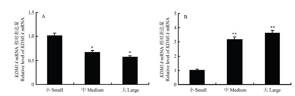
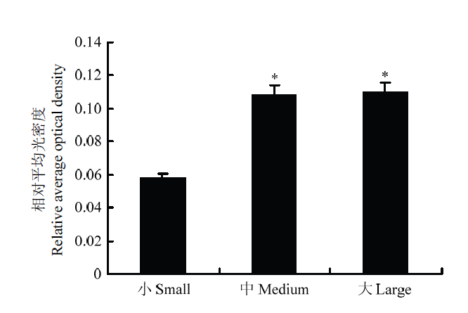
禹学礼, 昝林森, 邓雯, 庞有志, 王新庄 . 卵泡大小及卵泡液对牛卵母细胞体外受精后发育的影响. 中国农业科学, 2005,38(8):1664-1668.
|
|
YU X L, ZAN L S, DENG W, PANG Y Z, WANG X Z . Effects of follicle size and bovine follicular fluid on developmental competence of bovine oocytes following maturation, fertilization and culture in vitro. Scientia Agricultural Sinica, 2005,38(8):1664-1668. (in Chinese)
|
| [32] |
KIM J H, PARK U H, MOON M, UM S J, KIM E J . Negative regulation of ERa by a novel protein CAC1 through association with histone demethylase LSD1. Febs Letters, 2013,587(1):17-22.
|


 ),韩杰1,杨显英1,王艳1,王斌1,李键1(