


中国农业科学 ›› 2022, Vol. 55 ›› Issue (6): 1241-1252.doi: 10.3864/j.issn.0578-1752.2022.06.015
李文慧( ),贺依静,姜瑶,赵红宇,彭磊,李佳,芮荣,剧世强()
收稿日期:2021-01-29
接受日期:2021-06-17
出版日期:2022-03-16
发布日期:2022-03-25
通讯作者:
剧世强
作者简介:李文慧,E-mail: 基金资助:
LI WenHui(),HE YiJing,JIANG Yao,ZHAO HongYu,PENG Lei,LI Jia,RUI Rong,JU ShiQiang()
Received:2021-01-29
Accepted:2021-06-17
Online:2022-03-16
Published:2022-03-25
Contact:
ShiQiang JU
摘要:
【目的】探究伏马毒素B1(fumonisin B1,FB1)对猪卵母细胞体外成熟的影响及其潜在的作用机制, 为临床有效防治FB1所致的生殖毒性损伤提供理论参考。【方法】采集猪卵丘-卵母细胞复合体(cumulus oocyte complexes, COCs)进行随机分组,在体外成熟培养过程中分别用不同浓度FB1(0、10、20和30 μg·mL-1)处理44 h后,统计卵母细胞第一极体(first polar body, PB1)排出和激活后胚胎发育情况;通过免疫荧光染色结合共聚焦显微镜技术进一步检测FB1对卵母细胞减数分裂进程和细胞骨架结构的影响;为进一步探究FB1对猪卵母细胞毒性损伤的作用机制,分别采用JC-1、Annexin V-FITC和LC3A/B荧光染色检测各组卵母细胞内线粒体功能、早期凋亡和自噬水平,并在此基础上,进一步通过Western blotting分析了凋亡/自噬相关蛋白的表达情况。【结果】FB1处理对卵母细胞成熟具有明显的抑制作用,PB1排出率呈浓度依赖性下降,当FB1浓度达到20 μg·mL-1以上时,PB1排出率显著降低(P<0.01),并使卵母细胞孤雌激活后胚胎的卵裂率及囊胚率均显著降低(P<0.01),对卵母细胞的发育潜力有一定的损伤作用。细胞周期分析结果表明,FB1处理还会导致减数分裂周期进程紊乱,使阻滞在生发泡破裂期(germinal vesicle breakdown,GVBD)的卵母细胞比例显著升高(P<0.01)成功发育至第二次减数分裂中期(metaphase II,MII)细胞比例明显下降(P<0.01),同时,卵母细胞中纺锤体异常的比例显著升高(P<0.01)、胞膜上的微丝分布显著减少(P<0.05)。进一步的研究结果表明,与对照组相比,FB1处理组卵母细胞线粒体膜电位明显降低(P<0.05),线粒体功能受损,同时,FB1处理组卵母细胞早期凋亡率显著增加(P<0.01),细胞自噬水平也显著升高(P<0.01)。Western blotting分析结果显示,FB1处理组卵母细胞中促凋亡蛋白BAX和自噬蛋白LC3A/B II的表达均显著上调(P<0.05),抑凋亡蛋白BCL2的表达显著下调(P<0.05),提示早期凋亡和自噬的发生。【结论】FB1对猪卵母细胞体外成熟及其激活后胚胎发育具有明显的毒性损伤作用,致使减数分裂周期阻滞,纺锤体结构紊乱、微丝分布减少和线粒体损伤,其毒性作用机制与诱导卵母细胞凋亡和自噬有关。
李文慧,贺依静,姜瑶,赵红宇,彭磊,李佳,芮荣,剧世强. 伏马毒素B1对猪体外成熟卵母细胞凋亡与自噬的影响[J]. 中国农业科学, 2022, 55(6): 1241-1252.
LI WenHui,HE YiJing,JIANG Yao,ZHAO HongYu,PENG Lei,LI Jia,RUI Rong,JU ShiQiang. Effects of FB1 on Apoptosis and Autophagy of Porcine Oocytes in vitro Maturation[J]. Scientia Agricultura Sinica, 2022, 55(6): 1241-1252.

图1
不同浓度FB1对猪卵母细胞第一极体排出率的影响 A:卵母细胞体外培养44 h后的形态(黑色箭头指向第一极体);B:不同浓度FB1处理对PB1排出率的影响"
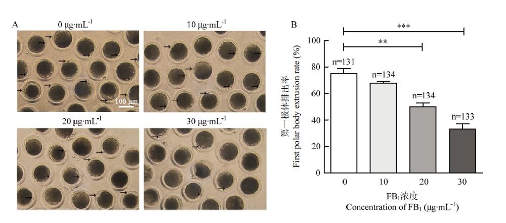
图2
FB1处理对卵母细胞激活后胚胎卵裂率和囊胚发育率的影响 A:2细胞/4细胞胚胎和囊胚的形态(黑色箭头指向卵裂的胚胎);B:卵裂率;C:囊胚率"
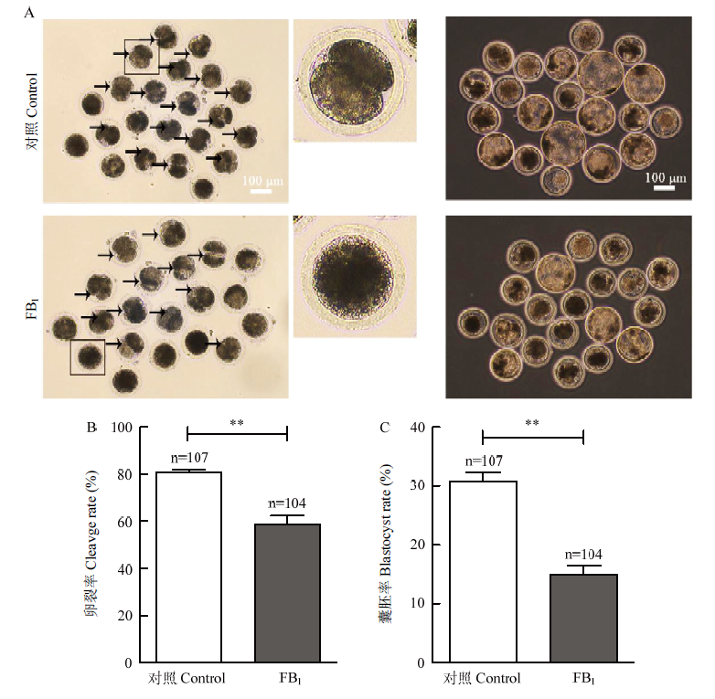

图3
猪卵母细胞减数分裂过程中的染色体和微管的动态分布典型图像 蓝色:染色体;绿色:微管"
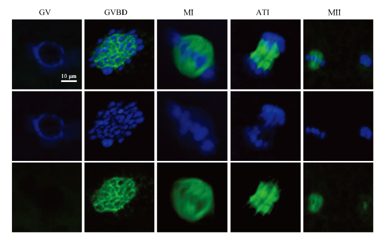
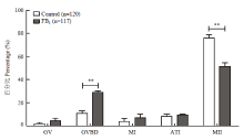
图4
FB1对猪卵母细胞减数分裂过程中细胞周期进程(44 h)的影响 GV:生发泡期;GVBD:生发泡破裂期;MI:第一次减数分裂中期;ATI:第一次减数分裂后-末期;MII:第二次减数分裂中期"
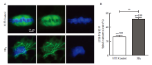
图5
FB1对猪卵母细胞纺锤体结构的影响 A:纺锤体结构和染色体排列的代表性图像(蓝色:染色体;绿色:微管);B:纺锤体异常百分比"
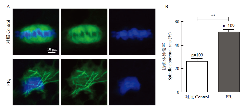
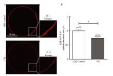
图6
FB1对猪卵母细胞微丝表达及分布的影响 A:微丝分布的代表性图像(红色:微丝);B:胞膜上微丝的相对平均荧光强度"
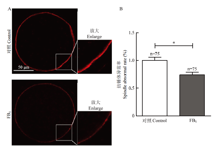

图7
FB1对猪卵母细胞线粒体膜电位的影响 A:JC-1染色的代表性图像(绿色:JC-1单体;红色:JC-1聚集体;黄色:合并图);B:JC-1红/绿信号的相对平均荧光强度"
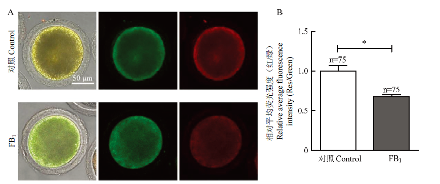

图8
FB1对猪卵母细胞早期凋亡率和凋亡相关蛋白表达的影响 A:Annexin-V-FITC染色的代表性图像和早期凋亡率(绿色:Annexin V荧光信号);B:BAX和BCL2蛋白表达"
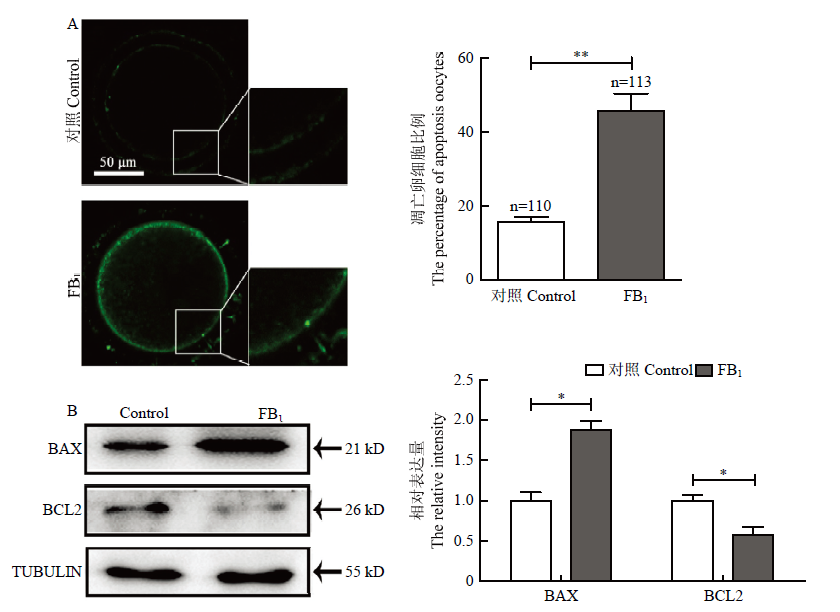
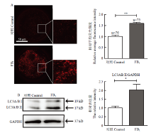
图9
FB1对猪卵母细胞自噬标记物LC3A/B蛋白表达的影响 A:LC3A/B染色的代表性图像和LC3A/B信号的相对平均荧光强度(红色:LC3荧光信号);B:LC3A/B蛋白表达"

| [39] |
YIN S T, GUO X, LI J H, FAN L H, HU H B. Fumonisin B1 induces autophagic cell death via activation of ERN1-MAPK8/9/10 pathway in monkey kidney MARC-145 cells. Archives of Toxicology, 2016,90(4):985-996. doi: 10.1007/s00204-015-1514-9.
doi: 10.1007/s00204-015-1514-9 |
| [40] |
ZHANG H, DIAO X, LI N, LIU C L. FB1-induced programmed cell death in hemocytes of Ostrinia furnacalis. Toxicon, 2018,146:114-119. doi: 10.1016/j.toxicon.2018.02.052.
doi: 10.1016/j.toxicon.2018.02.052 |
| [41] |
KHAN R B, PHULUKDAREE A, CHUTURGOON A A. Fumonisin B1 induces oxidative stress in oesophageal (SNO) cancer cells. Toxicon, 2018,141:104-111. doi: 10.1016/j.toxicon.2017.12.041.
doi: 10.1016/j.toxicon.2017.12.041 |
| [42] |
CHEN J, YANG S H, HUANG S, YAN R, WANG M Y, CHEN S, CAI J, LONG M, LI P. Transcriptome study reveals apoptosis of porcine kidney cells induced by fumonisin B1 via TNF signalling pathway. Food and Chemical Toxicology, 2020,139:111274. doi: 10.1016/j.fct.2020.111274.
doi: 10.1016/j.fct.2020.111274 |
| [43] |
D'ORSI B, MATEYKA J, PREHN J H M. Control of mitochondrial physiology and cell death by the Bcl-2 family proteins Bax and Bok. Neurochemistry International, 2017,109:162-170. doi: 10.1016/j.neuint.2017.03.010.
doi: 10.1016/j.neuint.2017.03.010 |
| [44] |
DENTON D, XU T Q, KUMAR S. Autophagy as a pro-death pathway. Immunology and Cell Biology, 2015,93(1):35-42. doi: 10.1038/icb.2014.85.
doi: 10.1038/icb.2014.85 |
| [1] |
KAMLE M, MAHATO D K, DEVI S, LEE K E, KANG S G, KUMAR P. Fumonisins: impact on agriculture, food, and human health and their management strategies. Toxins, 2019,11(6):328. doi: 10.3390/toxins11060328.
doi: 10.3390/toxins11060328 |
| [2] |
RHEEDER J P, MARASAS W F O, VISMER H F. Production of fumonisin analogs by Fusarium species. Applied and Environmental Microbiology, 2002,68(5):2101-2105. doi: 10.1128/AEM.68.5.2101-2105.2002.
doi: 10.1128/AEM.68.5.2101-2105.2002 |
| [3] |
ARUMUGAM T, GHAZI T, CHUTURGOON A. Fumonisin B1 epigenetically regulates PTEN expression and modulates DNA damage checkpoint regulation in HepG2 liver cells. Toxins, 2020,12(10):625. doi: 10.3390/toxins12100625.
doi: 10.3390/toxins12100625 |
| [4] |
STOEV S D, GUNDASHEVA D, ZARKOV I, MIRCHEVA T, ZAPRYANOVA D, DENEV S, MITEV Y, DASKALOV H, DUTTON M, MWANZA M, SCHNEIDER Y J. Experimental mycotoxic nephropathy in pigs provoked by a mouldy diet containing ochratoxin A and fumonisin B1. Experimental and Toxicologic Pathology, 2012,64(7/8):733-741. doi: 10.1016/j.etp.2011.01.008.
doi: 10.1016/j.etp.2011.01.008 |
| [5] |
STOCKMANN-JUVALA H, ALENIUS H, SAVOLAINEN K. Effects of fumonisin B(1) on the expression of cytokines and chemokines in human dendritic cells. Food and Chemical Toxicology, 2008,46(5):1444-1451. doi: 10.1016/j.fct.2007.12.004.
doi: 10.1016/j.fct.2007.12.004 |
| [6] |
DOMIJAN A M. Fumonisin B(1): a neurotoxic mycotoxin. Arhiv za higijenu rada i toksikologiju. 2012,63(4):531-544. doi: 10.2478/10004-1254-63-2012-2239.
doi: 10.2478/10004-1254-63-2012-2239 |
| [45] |
FULDA S, KÖGEL D. Cell death by autophagy: emerging molecular mechanisms and implications for cancer therapy. Oncogene, 2015,34(40):5105-5113. doi: 10.1038/onc.2014.458.
doi: 10.1038/onc.2014.458 |
| [7] |
GBORE F A. Brain and hypophyseal acetylcholinesterase activity of pubertal boars fed dietary fumonisin B1. Journal of Animal Physiology and Animal Nutrition, 2010,94(5):e123-e129. doi: 10.1111/j.1439-0396.2010.00992.x.
doi: 10.1111/j.1439-0396.2010.00992.x |
| [8] |
LUMSANGKUL C, CHIANG H I, LO N W, FAN Y K, JU J C. Developmental toxicity of mycotoxin fumonisin B1 in animal embryogenesis: an overview. Toxins, 2019,11(2):114. doi: 10.3390/toxins11020114.
doi: 10.3390/toxins11020114 |
| [9] |
SUN G J, WANG S K, HU X, SU J J, HUANG T R, YU J H, TANG L L, GAO W M, WANG J S. Fumonisin B1 contamination of home- grown corn in high-risk areas for esophageal and liver cancer in China. Food Additives & Contaminants, 2007,24(2):181-185. doi: 10.1080/02652030601013471.
doi: 10.1080/02652030601013471 |
| [10] |
LIU X Y, FAN L H, YIN S T, CHEN H, HU H B. Molecular mechanisms of fumonisin B1-induced toxicities and its applications in the mechanism-based interventions. Toxicon, 2019,167:1-5. doi: 10.1016/j.toxicon.2019.06.009.
doi: 10.1016/j.toxicon.2019.06.009 |
| [11] |
DESAI K N, SULLARDS M C, ALLEGOOD J, WANG E, SCHMELZ E M, HARTL M, HUMPF H U, LIOTTA D C, PENG Q, MERRILL A H Jr. Fumonisins and fumonisin analogs as inhibitors of ceramide synthase and inducers of apoptosis. Biochimica et Biophysica Acta (BBA) - Molecular and Cell Biology of Lipids, 2002,1585(2/3):188-192. doi: 10.1016/S1388-1981(02)00340-2.
doi: 10.1016/S1388-1981(02)00340-2 |
| [12] |
STOCKMANN-JUVALA H, MIKKOLA J, NAARALA J, LOIKKANEN J, ELOVAARA E, SAVOLAINEN K. Oxidative stress induced by fumonisin B1 in continuous human and rodent neural cell cultures. Free Radical Research, 2004,38(9):933-942. doi: 10.1080/10715760412331273205.
doi: 10.1080/10715760412331273205 |
| [13] |
KIM S H, SINGH M P, SHARMA C, KANG S C. Fumonisin B1 actuates oxidative stress-associated colonic damage via apoptosis and autophagy activation in murine model. Journal of Biochemical and Molecular Toxicology, 2018,32(7):e22161. doi: 10.1002/jbt.22161.
doi: 10.1002/jbt.22161 |
| [14] |
GBORE F A. Reproductive organ weights and semen quality of pubertal boars fed dietary fumonisin B1. Animal, 2009,3(8):1133-1137. doi: 10.1017/S1751731109004467.
doi: 10.1017/S1751731109004467 |
| [15] |
HENRY M H, WYATT R D. The toxicity of fumonisin B1, B2, and B3, individually and in combination, in chicken embryos. Poultry Science, 2001,80(4):401-407. doi: 10.1093/ps/80.4.401.
doi: 10.1093/ps/80.4.401 |
| [16] |
CORTINOVIS C, PIZZO F, SPICER L J, CALONI F. Fusarium mycotoxins: effects on reproductive function in domestic animals—A review. Theriogenology, 2013,80(6):557-564. doi: 10.1016/j.theriogenology.2013.06.018.
doi: 10.1016/j.theriogenology.2013.06.018 |
| [17] | 郭隽, 张立实, 彭双清. 镰刀菌毒素生殖发育毒性研究进展. 中国食品卫生杂志, 2013(5):474-478. |
| GUO J, ZHANG L S, PENG S Q. Review of reproductive and developmental toxicity studies of Fusarium toxins. Chinese Journal of Food Hygiene, 2013(5):474-478. (in Chinese) | |
| [18] |
SOMOSKŐI B, KOVÁCS M, CSEH S. Effects of T-2 and Fumonisin B1 combined treatment on in vitro mouse embryo development and blastocyst quality. Toxicology and Industrial Health, 2018,34(5):353-360. doi: 10.1177/0748233718764039.
doi: 10.1177/0748233718764039 |
| [19] |
CORTINOVIS C, CALONI F, SCHREIBER N B, SPICER L J. Effects of fumonisin B1 alone and combined with deoxynivalenol or Zearalenone on porcine granulosa cell proliferation and steroid production. Theriogenology, 2014,81(8):1042-1049. doi: 10.1016/j.theriogenology.2014.01.027.
doi: 10.1016/j.theriogenology.2014.01.027 |
| [20] |
SHI F Y, LI W H, ZHAO H Y, HE Y J, JIANG Y, NI J, ABBASI B, RUI R, JU S Q. Microcystin-LR exposure results in aberrant spindles and induces apoptosis in porcine oocytes. Theriogenology, 2020,158:358-367. doi: 10.1016/j.theriogenology.2020.09.031.
doi: 10.1016/j.theriogenology.2020.09.031 |
| [21] |
CUI P P, ABBASI B, LIN D F, RUI R, JU S Q. Aurora A inhibition disrupts chromosome condensation and spindle assembly during the first embryonic division in pigs. Reproduction in Domestic Animals, 2020,55(5):584-593. doi: 10.1111/rda.13655.
doi: 10.1111/rda.13655 |
| [22] |
YANG C X, WANG P C, LIU S, MIAO J K, LIU X M, MIAO Y L, DU Z Q. Long noncoding RNA 2193 regulates meiosis through global epigenetic modification and cytoskeleton organization in pig oocytes. Journal of Cellular Physiology, 2020,235(11):8304-8318. doi: 10.1002/jcp.29675.
doi: 10.1002/jcp.29675 |
| [23] |
DING Z M, AHMAD M J, MENG F, CHEN F, WANG Y S, ZHAO X Z, ZHANG S X, MIAO Y L, XIONG J J, HUO L J. Triclocarban exposure affects mouse oocyte in vitro maturation through inducing mitochondrial dysfunction and oxidative stress. Environmental Pollution, 2020,262:114271. doi: 10.1016/j.envpol.2020.114271.
doi: 10.1016/j.envpol.2020.114271 |
| [24] |
MAN W R, GU J, WANG B, ZHANG M M, HU J Q, LIN J, SUN D, XIONG Z Y, GU X M, HAO K K, GUO B L, WEI G L, ZHANG L, SONG R, LI C Y, WANG H C, SUN D D. SHANK3 co-ordinately regulates autophagy and apoptosis in myocardial infarction. Frontiers in Physiology, 2020,11:1082. doi: 10.3389/fphys.2020.01082.
doi: 10.3389/fphys.2020.01082 |
| [25] |
REYES J M, ROSS P J. Cytoplasmic polyadenylation in mammalian oocyte maturation. Wiley Interdisciplinary Reviews: RNA, 2016,7(1):71-89. doi: 10.1002/wrna.1316.
doi: 10.1002/wrna.1316 |
| [26] |
黄向月, 熊显荣, 韩杰, 杨显英, 王艳, 王斌, 李键. KDM1A在牦牛卵泡发育过程中的表达. 中国农业科学, 2019,52(24):4624-4631. doi: 10.3864/j.issn.0578-1752.2019.24.016.
doi: 10.3864/j.issn.0578-1752.2019.24.016 |
|
HUANG X Y, XIONG X R, HAN J, YANG X Y, WANG Y, WANG B, LI J. Expression pattern of KDM1A in the development of yak follicles. Scientia Agricultura Sinica, 2019,52(24):4624-4631. doi: 10.3864/j.issn.0578-1752.2019.24.016. (in Chinese)
doi: 10.3864/j.issn.0578-1752.2019.24.016 |
|
| [27] |
BRUNET S, MARO B. Cytoskeleton and cell cycle control during meiotic maturation of the mouse oocyte: integrating time and space. Reproduction (Cambridge, England), 2005,130(6):801-811. doi: 10.1530/rep.1.00364.
doi: 10.1530/rep.1.00364 |
| [28] |
ZHANG H, ZHANG L Y, DIAO X, LI N, LIU C L. Toxicity of the mycotoxin fumonisin B1 on the insect Sf9 cell line. Toxicon, 2017,129:20-27. doi: 10.1016/j.toxicon.2017.01.018.
doi: 10.1016/j.toxicon.2017.01.018 |
| [29] |
MARIN D E, GOUZE M E, TARANU I, OSWALD I P. Fumonisin B1 alters cell cycle progression and interleukin-2 synthesis in swine peripheral blood mononuclear cells. Molecular Nutrition & Food Research, 2007,51(11):1406-1412. doi: 10.1002/mnfr.200700131.
doi: 10.1002/mnfr.200700131 |
| [30] | 秦伟森. 伏马菌素FB1对人脐静脉血管内皮细胞的毒性作用研究[D]. 广州: 华南农业大学, 2016. |
| QIN W S. Cellular and relevant factors of toxicity of fumonisin B1 in human umbilical vein endothelial cells[D]. Guangzhou: South China Agricultural University, 2016. (in Chinese) | |
| [31] |
SUN S C, KIM N H. Molecular mechanisms of asymmetric division in oocytes. Microscopy and Microanalysis, 2013,19(4):883-897. doi: 10.1017/S1431927613001566.
doi: 10.1017/S1431927613001566 |
| [32] |
ZHAO X, WANG Y, LIU J L, ZHANG J H, ZHANG S C, OUYANG Y, HUANG J T, PENG X Y, ZENG Z, HU Z Q. Fumonisin B1 affects the biophysical properties, migration and cytoskeletal structure of human umbilical vein endothelial cells. Cell Biochemistry and Biophysics, 2020,78(3):375-382. doi: 10.1007/s12013-020-00923-4.
doi: 10.1007/s12013-020-00923-4 |
| [33] |
AL-ZUBAIDI U, LIU J, CINAR O, ROBKER R L, ADHIKARI D, CARROLL J. The spatio-temporal dynamics of mitochondrial membrane potential during oocyte maturation. Molecular Human Reproduction, 2019,25(11):695-705. doi: 10.1093/molehr/gaz055.
doi: 10.1093/molehr/gaz055 |
| [34] |
BOCK F J, TAIT S W G. Mitochondria as multifaceted regulators of cell death. Nature Reviews Molecular Cell Biology, 2020,21(2):85-100. doi: 10.1038/s41580-019-0173-8.
doi: 10.1038/s41580-019-0173-8 |
| [35] |
TARAZONA A, RODRÍGUEZ J, RESTREPO L, OLIVERA-ANGEL M. Mitochondrial activity, distribution and segregation in bovine oocytes and in embryos produced in vitro. Reproduction in Domestic Animals, 2006,41(1):5-11. doi: 10.1111/j.1439-0531.2006.00615.x.
doi: 10.1111/j.1439-0531.2006.00615.x |
| [36] |
SHEIK ABDUL N, MARNEWICK J L. Fumonisin B1-induced mitochondrial toxicity and hepatoprotective potential of rooibos: an update. Journal of Applied Toxicology, 2020,40(12):1602-1613. doi: 10.1002/jat.4036.
doi: 10.1002/jat.4036 |
| [37] |
ARUMUGAM T, PILLAY Y, GHAZI T, NAGIAH S, ABDUL N S, CHUTURGOON A A. Fumonisin B1-induced oxidative stress triggers Nrf2-mediated antioxidant response in human hepatocellular carcinoma (HepG2) cells. Mycotoxin Research, 2019,35(1):99-109. doi: 10.1007/s12550-018-0335-0.
doi: 10.1007/s12550-018-0335-0 |
| [38] |
潘阳阳, 李秦, 崔燕, 樊江峰, 杨琨, 何俊峰, 余四九. EGF、EGFR在牦牛卵母细胞中的表达及对胚胎发育能力的作用. 中国农业科学, 2015,48(12):2439-2448. doi: 10.3864/j.issn.0578-1752.2015.12.017.
doi: 10.3864/j.issn.0578-1752.2015.12.017 |
|
PAN Y Y, LI Q, CUI Y, FAN J F, YANG K, HE J F, YU S J. The expression of EGF and EGFR in yak oocyte and its function on development competence of embryo. Scientia Agricultura Sinica, 2015,48(12):2439-2448. doi: 10.3864/j.issn.0578-1752.2015.12.017. (in Chinese)
doi: 10.3864/j.issn.0578-1752.2015.12.017 |
| [1] | 刘玉芳,陈玉林,周祖阳,储明星. miR-221-3p靶向BCL2L11调控小尾寒羊卵泡颗粒细胞凋亡[J]. 中国农业科学, 2022, 55(9): 1868-1876. |
| [2] | 阿依木古丽·阿不都热依木,阿尔祖古丽·阿依丁,王家敏,石嘉琛,马芳芳,蔡勇,乔自林. 大豆异黄酮对牦牛卵巢颗粒细胞增殖和凋亡的影响[J]. 中国农业科学, 2022, 55(8): 1667-1675. |
| [3] | 邢明杰,顾宪红,王枭鸿,郝月. IL-15过表达对猪骨骼肌细胞成肌分化的影响[J]. 中国农业科学, 2022, 55(18): 3652-3663. |
| [4] | 杨昌沛,王乃秀,汪锴,黄子晴,林海烂,张莉,张晨,冯露秋,甘玲. 外源性γ-氨基丁酸抵抗仔猪氧化应激的效果及机制[J]. 中国农业科学, 2022, 55(17): 3437-3449. |
| [5] | 冯云奎,王健,马金亮,张柳明,李拥军. miR-31-5p对山羊毛囊干细胞增殖和凋亡的影响[J]. 中国农业科学, 2021, 54(23): 5132-5143. |
| [6] | 马梦楠,王慧明,王苗苗,姚望,张金璧,潘增祥. 猪卵泡闭锁过程中circINHBB的鉴定及其对颗粒细胞凋亡的影响[J]. 中国农业科学, 2021, 54(18): 3998-4007. |
| [7] | 李闰婷,陈龙欣,张丽萌,何海迎,王泳,杨若晨,段春辉,刘月琴,王玉琴,张英杰. 粒细胞集落刺激因子在羊成纤维细胞中的表达及对细胞增殖和凋亡的影响[J]. 中国农业科学, 2021, 54(11): 2434-2444. |
| [8] | 黄峰,魏起超,李侠,刘春梅,张春晖. 细胞凋亡对宰后肌肉嫩化作用机理的研究进展[J]. 中国农业科学, 2021, 54(10): 2192-2202. |
| [9] | 张鑫,霍孔林,宋星星,张多妮,胡文,胡传活,李珣. GnIH通过p38MAPK信号通路对猪卵巢颗粒细胞自噬与凋亡的影响[J]. 中国农业科学, 2020, 53(9): 1904-1912. |
| [10] | 李桢,杨世雄,牛胜,张宁,李欣,张阳阳,贾云飞,田志雄,宁官保,张鼎,田文霞. 重组GSTA3蛋白对福美双诱导的肉鸡TD中抗凋亡基因BAG-3表达的影响[J]. 中国农业科学, 2020, 53(9): 1921-1930. |
| [11] | 潘阳阳,王萌,芮弦,王立斌,何翃闳,王靖雷,马睿,徐庚全,崔燕,樊江峰,余四九. IGF-1调控RBM3表达抑制低温应激诱导牦牛卵丘细胞凋亡[J]. 中国农业科学, 2020, 53(11): 2285-2296. |
| [12] | 栗永华, 车路平, 仇旭升, 谭磊, 孙英杰, 刘炜炜, 宋翠萍, 廖瑛, 丁铲, 王金泉, 孟春春. 鸡TIGAR基因真核表达质粒的构建及其抗凋亡功能评价[J]. 中国农业科学, 2019, 52(6): 1102-1109. |
| [13] | 陈鹏,包希艳,康涛涛,董战旗,朱艳,潘敏慧,鲁成. 家蚕IAP互作蛋白的筛选、鉴定及其对BmNPV增殖的影响[J]. 中国农业科学, 2019, 52(3): 558-567. |
| [14] | 邓少锋,叶佐东,范双旗,陈金顶,张静远,朱梦娇,赵明秋. PK-15细胞中与CSFV感染相关的microRNAs筛选及 miR-214的功能研究[J]. 中国农业科学, 2018, 51(21): 4157-4168. |
| [15] | 陈琳琳,侯莹,丁胜利,施艳,李洪连. 假禾谷镰孢细胞凋亡基因FpTatD的鉴定与表达分析[J]. 中国农业科学, 2016, 49(12): 2301-2309. |
|
||