


中国农业科学 ›› 2026, Vol. 59 ›› Issue (6): 1333-1347.doi: 10.3864/j.issn.0578-1752.2026.06.014
格日其木格1( ), 普布占堆2, 徐青1(), 侯玲玲1()
收稿日期:2025-10-17
接受日期:2025-12-18
出版日期:2026-03-16
发布日期:2026-03-24
通信作者:
联系方式:
格日其木格,E-mail:2975747682@qq.com。
基金资助:
GERIQIMUGE1(), PUBUZHANDUI2, XU Qing1(), HOU LingLing1()
Received:2025-10-17
Accepted:2025-12-18
Published:2026-03-16
Online:2026-03-24
摘要:
【背景】低氧是一种独特的环境应激,广泛地影响着呼吸、循环、泌尿、消化等多个系统的生理功能,在个体、组织器官以及细胞水平产生不同程度的影响,严重时引起机体组织器官损伤和疾病,在细胞水平涉及形态结构、代谢、增殖、自噬及凋亡等多个方面。肾脏是泌尿系统的重要组成部分,对于维持体内平衡、调节水电解质和酸碱平衡以及消除代谢废物至关重要,属于低氧敏感组织。目前,低氧对牛肾脏细胞的影响及其分子机制尚不明确。【目的】以牛肾细胞系(madin-darby bovine kidney,MDBK)为细胞模型,探讨不同低氧浓度(11% O2、5% O2和1% O2)和不同低氧处理时间(24、48和72 h)对牛肾细胞的增殖能力、超微结构、线粒体功能及低氧应激能力的影响,为进一步探究牛肾脏低氧适应机制提供了试验依据,也为深入理解低氧条件下细胞的生存策略和线粒体自噬的调控机制提供基础。【方法】在体内,不同细胞和细胞器感受到的氧分压不同,在常氧下,氧分压为159.22 mmHg(20.95%),动脉氧分压为100 mmHg(13%),静脉氧分压(PaO2)为40 mmHg(5.2%),线粒体氧分压为4—20 mmHg(0.52%—2.60%),鉴于此,本研究选择11% O2、5% O2和1% O2浓度作为低氧处理条件。将牛肾细胞系MDBK接种于含10%胎牛血清的DMEM完全培养基中,在37 ℃、5% CO2条件下培养至对数生长期,随后转移至三气培养箱,分别在11% O2、5% O2和1% O2浓度下培养24、48和72 h,通过MTT法检测低氧对牛肾细胞增殖能力的影响,通过透射扫描电子显微镜观察低氧条件下MDBK细胞超微结构的变化,通过JC-1荧光探针法检测线粒体膜电位,通过DCFH-DA荧光探针法检测活性氧的产生,通过实时荧光定量PCR和Western Blot检测低氧相关基因以及自噬相关基因的表达情况。【结果】随着氧气浓度的下降和处理时间的延长,细胞增殖能力受到显著抑制,且氧气浓度越低处理时间越长抑制越显著;低氧导致线粒体结构损伤和功能障碍,表现为线粒体肿胀、嵴排列紊乱甚至消失,且线粒体数量也显著减少,部分线粒体发生固缩,出现线粒体被膜性结构包绕及线粒体自噬的现象,同时线粒体膜电位显著降低;细胞内活性氧水平显著升高;低氧相关基因EPAS1和PPARα的表达显著升高,线粒体自噬相关基因PINK1、PRKN、BNIP3和BNIP3L(NIX)的表达水平显著上调。【结论】5%和1%氧气浓度处理显著抑制牛肾细胞的增殖活力,并导致线粒体结构损伤和功能障碍,线粒体膜电位下降,活性氧产生增加。在应答低氧胁迫过程中,牛肾细胞通过激活线粒体自噬以清除功能失调的线粒体,从而减轻氧化应激,以维持细胞内环境的稳定。
格日其木格, 普布占堆, 徐青, 侯玲玲. 低氧对牛肾细胞增殖和线粒体自噬的影响[J]. 中国农业科学, 2026, 59(6): 1333-1347.
GERIQIMUGE, PUBUZHANDUI, XU Qing, HOU LingLing. Effects of Hypoxia on Proliferation of Bovine Renal Cells and Mitochondrial Autophagy[J]. Scientia Agricultura Sinica, 2026, 59(6): 1333-1347.
表1
细胞接种密度"
| 10 cm皿 10 cm dish (cells/dish) | 3.5 cm皿 3.5 cm dish (cells/dish) | 6孔板 6-well plate (cells/well) | 24孔板 24-well plate (cells/well) | 96孔板 96-well plate (cells/well) | |
|---|---|---|---|---|---|
| 24 h | 3×10⁶ | 4.8×10⁵ | 7.2×10⁵ | / | 7×10³ |
| 48 h | 2×10⁶ | 2.8×10⁵ | 3.6×10⁵ | / | 5×10³ |
| 72 h | 1.3×10⁶ | 1.6×10⁵ | 1.8×10⁵ | 4×10⁴ | 3×10³ |
表2
RT-qPCR反应体系"
| 成分 Component | 使用量 Volume (μL) |
|---|---|
| 2 × FastFire qPCR PreMix | 5 |
| 正向引物(10 μmol·L-1)Primer-Forward(10μmol·L-1) | 0.3 |
| 反向引物(10μmol·L-1)Primer-Reverse(10μmol·L-1) | 0.3 |
| cDNA模板 cDNA template | 1 |
| RNase-Free H2O | 3.4 |
| 合计 Total | 10 |
表3
RT-qPCR反应条件"
| 温度 Temperature (℃) | 时间 Time | 循环数 Cycle number |
|---|---|---|
| 95 | 1 min | 1 |
| 95 | 5 s | / |
| 60 | 30 s | 40 |
表4
qRT-PCR引物"
| 基因 Gene | 引物序列 Primer sequences (5'→3') | 产物大小 Product size (bp) | 退火温度 Annealing temperature ( ℃) |
|---|---|---|---|
| EPAS1 | F:AAGACATGTCCACAGAGCGG R:TGTCGTCGCAGTAGGTGAAC | 300 | 57.60 57.40 |
| PPARα | F:GCGGCCCCAGGTGGT R:GATGCTGGATGACTCCTCGG | 237 | 63.60 58.20 |
| LC3B | F:CCGACTTATCCGAGAGCAGC R:TGAGCTGTAAGCGCCTTCTT | 277 | 59.69 60.55 |
| BNIP3 | F:GAAGAACAGCTCCCAGTCCGA R:CCGACTGGACCAATCCCAA | 103 | 61.77 59.32 |
| NIX | F:ACTTAGAACAGCCGCCG R:CCACCCAGGAACTGTTGAGG | 97 | 55.40 58.70 |
| PINK1 | F:TGTGGAACATCTCGGCAGG R:GGACCTCCCTTGGGTCTTCT | 133 | 59.70 60.55 |
| PRKN | F:CAGTGGCCATGATAGAACCTGA R:CAGAATCGACCTCCACTGGG | 187 | 59.83 59.82 |
| MFN1 | F:AGACAGTTAATCAGCTGGCCC R:ATCTGTGCCCGGACTGTCTA | 140 | 52.38 55.00 |
| 18sRNA | F:GTAACCCGTTGAACCCCATT R:CCATCCAATCGGTAGTAGCG | 152 | 58.09 57.93 |
表5
浓缩胶和分离胶配制所需试剂"
| 试剂 Reagent | 15% 分离胶 15% resolving gel (mL) | 5% 浓缩胶 5% stacking gel (mL) |
|---|---|---|
| H2O | 2.3 | 5.5 |
| 30% 丙烯酰胺溶液 30% Acr-Bis | 5.0 | 1.3 |
| Tris-HCl | 2.5(pH 8.8,1.5 mol·L-1) | 1.0(pH 6.8,1.5 mol·L-1) |
| 10% 十二烷基硫酸钠 10% SDS | 0.1 | 0.08 |
| 10% 过硫酸铵 10% APS | 0.1 | 0.08 |
| 四甲基乙二胺 TEMED | 0.004 | 0.008 |
| 总计 Total | 10.0 | 8.0 |
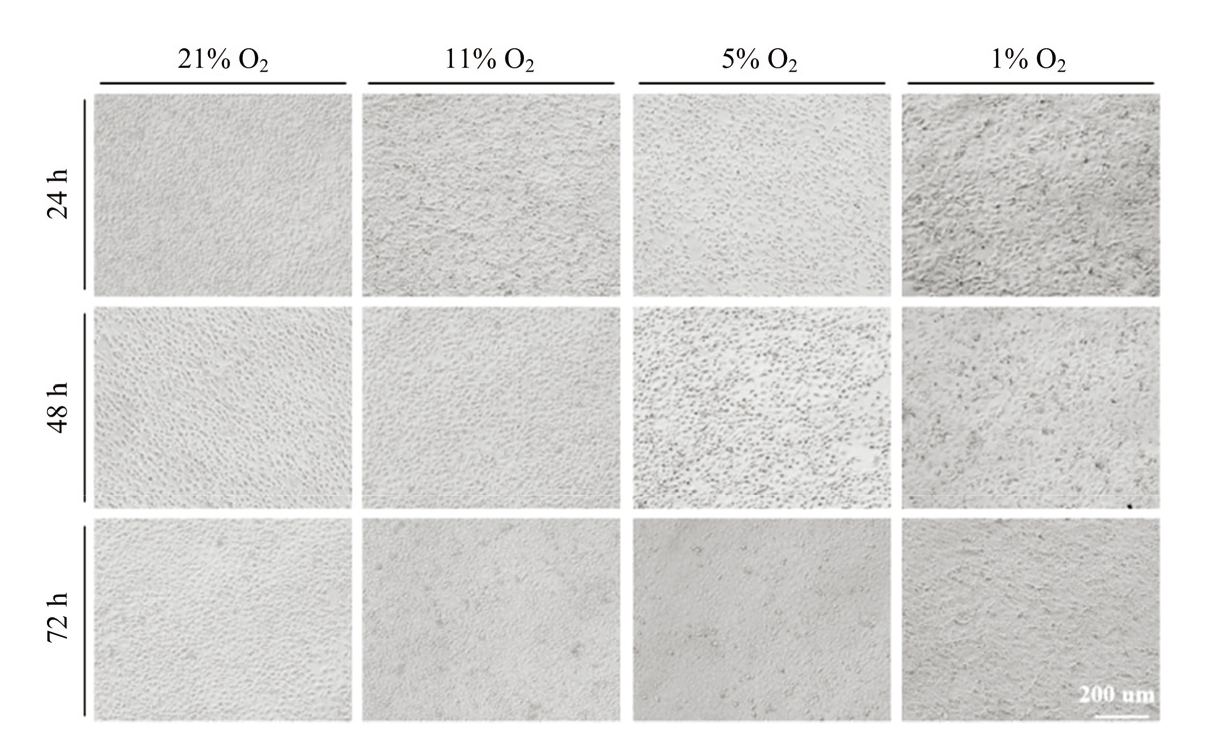
图1
不同低氧浓度对牛肾细胞系MDBK形态的影响"
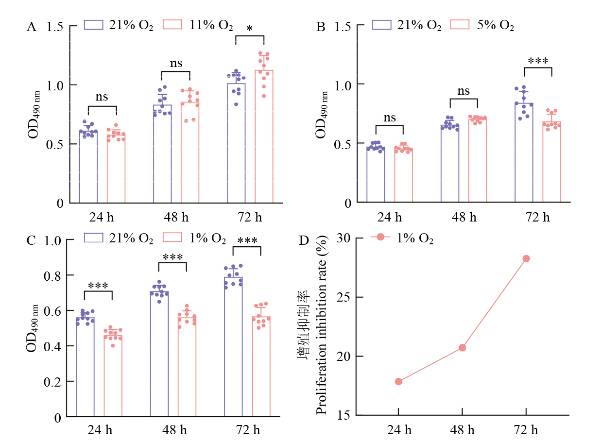
图2
MTT法检测不同氧气浓度和培养时间对牛肾细胞系MDBK增殖的影响 A—C:MTT法检测不同氧气浓度(21% O2、11% O2、5% O2和1% O2)及处理时间(24、48和72 h)下,牛肾细胞系MDBK增殖活力的变化,其中星号表示与对照组相比具有统计学显著性差异(*P < 0.05,**P< 0.01,***P < 0.001,ns表示无显著差异);D:“增殖抑制率”表示在1%O2浓度下处理24、48和72 h后, 牛肾细胞系MDBK增殖受到抑制的程度"

图3
5% O2和1% O2对牛肾细胞系MDBK超微结构的影响 透射电镜下观察到线粒体数量减少,线粒体固缩(绿色箭头标记),线粒体自噬(红色箭头标记)"
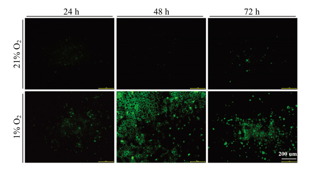
图4
荧光探针法检测牛肾细胞系MDBK中活性氧的产生"

图5
JC-1染色检测常氧和低氧条件下MDBK细胞线粒体膜电位变化 绿色荧光:JC-1单体(JC-1 monomers),线粒体受损;红色荧光:JC-1聚合体(JC-1 aggregates),线粒体正常"
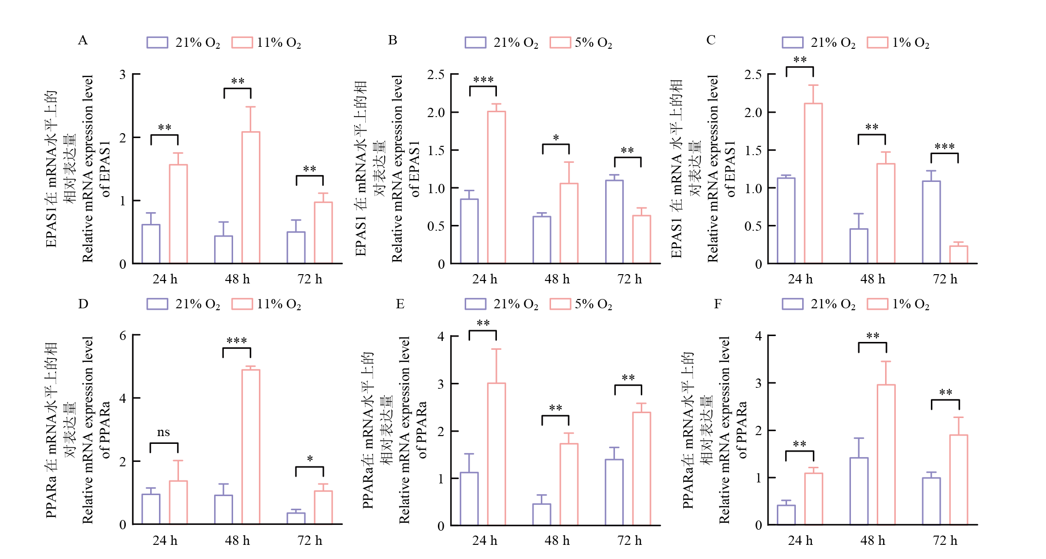
图6
低氧对牛肾细胞系MDBK低氧相关基因表达的影响 A—F:RT-qPCR检测不同氧气浓度(21% O2、11% O2、5% O2和1% O2)及处理时间(24、48和72 h)下,牛肾细胞系MDBK低氧相关基因EPAS1和PPARα的mRNA表达水平"
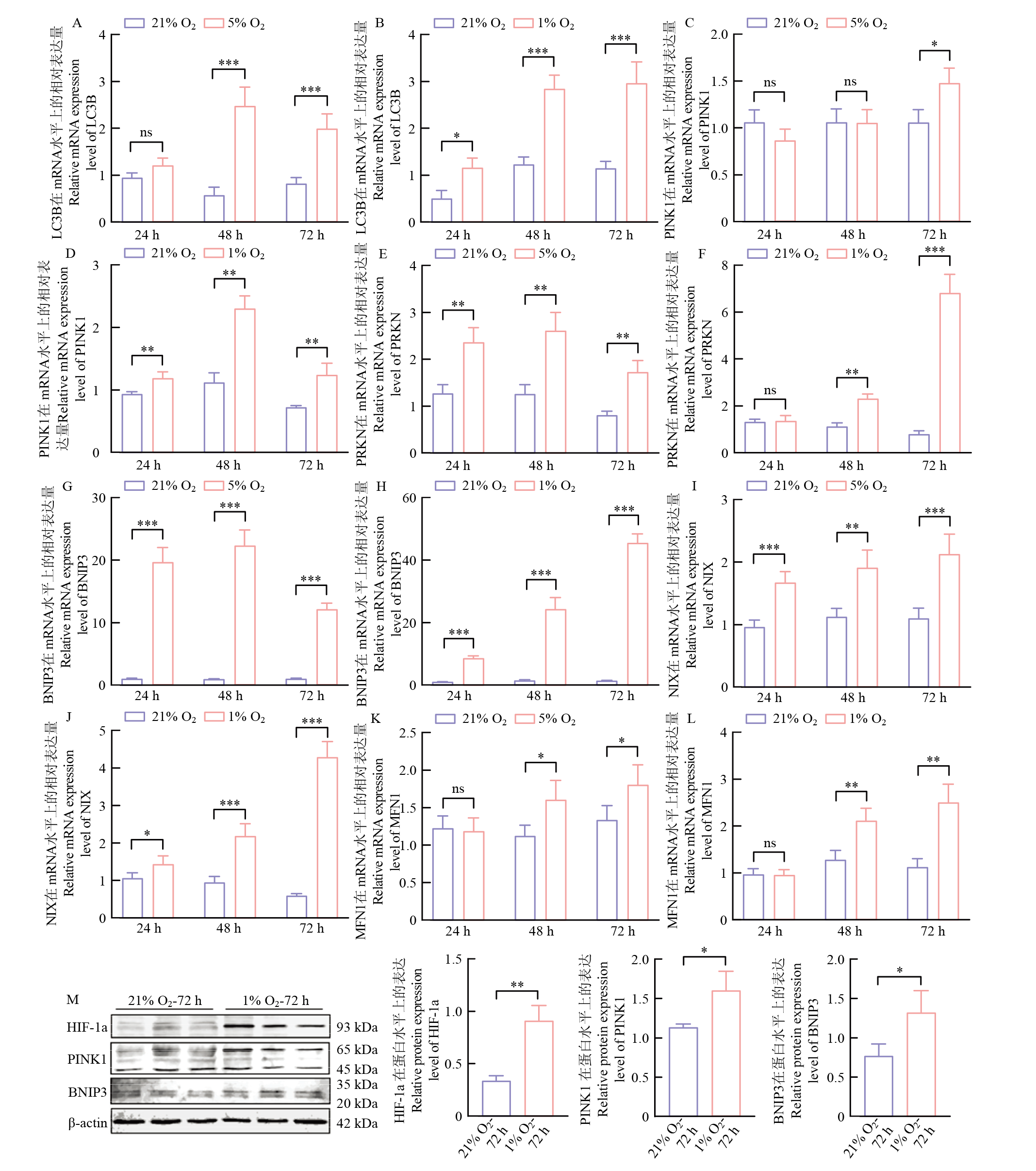
图7
低氧对牛肾细胞系MDBK自噬标记基因的影响 A—L:检测不同氧气浓度(21% O2、5% O2和1% O2)及处理时间(24、48和72 h)下,牛肾细胞系MDBK自噬标记基因LC3B、PINK1、PRKN、BNIP3、NIX和MFN1的mRNA水平上的表达。M:1% O2处理72 h后HIF-1a、PINK1和BNIP3蛋白水平上的表达"
| [1] |
doi: 10.3390/biom15040556 |
| [2] |
姚一凡, 张伊阳, 董仕慧, 李睿, 张兰, 周熳琳, 乔自林, 杨琨. 低氧下牦牛、黑白花牛肾间质成纤维细胞PDK1、Smad2、Caspase3等因子表达差异研究. 核农学报, 2023, 37(7): 1335-1343.
doi: 10.11869/j.issn.1000-8551.2023.07.1335 |
|
doi: 10.11869/j.issn.1000-8551.2023.07.1335 |
|
| [3] |
曾坐佳, 雷迁. 心肌高海拔低氧适应的研究进展. 实用医院临床杂志, 2023, 20(4): 194-196.
|
|
|
|
| [4] |
pmid: 31818357 |
| [5] |
张剑搏, 丁学智,
|
|
doi: 10.11843/j.issn.0366-6964.2019.09.001 |
|
| [6] |
doi: 10.3390/cells8030207 |
| [7] |
doi: 10.1016/j.ebiom.2022.103942 |
| [8] |
doi: 10.5713/ajas.14.0413 |
| [9] |
doi: 10.1093/gbe/evy264 pmid: 30517636 |
| [10] |
|
| [11] |
doi: 10.1016/j.ygeno.2024.110857 |
| [12] |
doi: 10.1007/s11325-018-1720-9 |
| [13] |
doi: 10.1016/j.ymthe.2022.08.013 |
| [14] |
doi: 10.3389/fendo.2021.626390 |
| [15] |
|
| [16] |
|
| [17] |
|
| [18] |
doi: 10.1016/j.arr.2022.101817 |
| [19] |
|
| [20] |
doi: 10.1038/s41467-020-18638-8 pmid: 33004791 |
| [21] |
doi: 10.1177/1933719118778798 pmid: 29848205 |
| [22] |
doi: 10.1016/j.bcp.2022.115350 |
| [23] |
doi: 10.1007/s00018-014-1645-9 pmid: 24858415 |
| [24] |
doi: 10.3390/ijms24032788 |
| [25] |
doi: 10.1186/1471-2121-11-11 pmid: 20109207 |
| [26] |
doi: 10.3390/ijms19123893 |
| [27] |
邵川, 鲁沈源, 励雯静, 隆玄, 李善群, 白春学. 慢性间歇低氧对大鼠肾脏组织细胞形态和超微结构的影响. 中国老年学杂志, 2011, 31(21): 4175-4177.
|
|
|
|
| [28] |
张勤文, 俞红贤, 荆海霞, 李莉, 魏青, 牛海林, 薛乾, 梁林. 基于骨骼肌线粒体超微结构研究生长期牦牛低氧适应性. 畜牧兽医学报, 2013, 44(3): 447-452.
|
|
|
|
| [29] |
徐媛媛, 黄丽清, 陆杏蓉, 冯超, 尚江华. β-巯基乙醇对雷帕霉素诱导的水牛颗粒细胞自噬、凋亡和类固醇激素分泌的影响. 中国农业科学, 2025, 58(11): 2265-2274. doi: 10.3864/j.issn.0578-1752.2025.11.014.
|
|
|
|
| [30] |
doi: 10.1016/j.bioactmat.2022.05.006 |
| [31] |
doi: 10.1080/15548627.2022.2084862 |
| [32] |
doi: 10.1152/ajpheart.01271.2010 |
| [33] |
doi: 10.1016/j.redox.2021.102047 |
| [34] |
doi: 10.1080/15548627.2020.1797280 |
| [35] |
doi: 10.7150/ijbs.80775 |
| [36] |
doi: 10.3389/fimmu.2025.1549276 |
| [37] |
|
| [38] |
doi: 10.3390/ani11082344 |
| [39] |
doi: 10.31083/j.fbl2902068 |
| [40] |
doi: 10.7150/ijms.88216 |
| [41] |
doi: 10.1152/physiol.00043.2024 |
| [42] |
doi: 10.1038/s41390-022-02193-7 |
| [1] | 岳潇雨, 赵世琛, 王勤. miR-362-3p靶向BMPR2调控马卵泡颗粒细胞增殖和类固醇激素合成[J]. 中国农业科学, 2026, 59(7): 1564-1575. |
| [2] | 王薇, 罗春海, 贾红豆, 刘佳金, 李丹阳, 付世新. 谷氨酰胺对氧化应激状态下胎衣不下奶牛内质网应激的影响[J]. 中国农业科学, 2025, 58(7): 1451-1462. |
| [3] | 王妞, 史昕冉, 张卫东, 王昕. FGF5和FGF21对绒山羊毛乳头细胞增殖的影响[J]. 中国农业科学, 2025, 58(4): 819-830. |
| [4] | 李蕊彤, 陈清梅, 徐建芳, 张军民, 司玮, 张铁鹰. 基于猪肠道类器官模型的甘草次酸抗氧化功能及机制研究[J]. 中国农业科学, 2025, 58(22): 4771-4785. |
| [5] | 李翔宇, 刘健茁, 胡丹丹, 刘耕瑜, 陈良宇, 李冰, 杜万里, 宋波. 玉米种质资源对瘤黑粉病的抗性评价及生理差异分析[J]. 中国农业科学, 2025, 58(13): 2504-2521. |
| [6] | 姜超, 张久盘, 宋雅萍, 宋小雨, 吴昊, 魏大为. FoxO1对牛骨骼肌细胞增殖、凋亡和分化的调控[J]. 中国农业科学, 2024, 57(6): 1191-1203. |
| [7] | 刘卓琳, 刘红云. 基于网络药理学和分子对接探究芹菜素缓解奶牛热应激及低氧应激的潜力与机制[J]. 中国农业科学, 2024, 57(5): 1010-1022. |
| [8] | 李凯利, 魏云晓, 种智力, 孟志刚, 王远, 梁成真, 陈全家, 张锐. 红蓝光促进陆地棉愈伤组织诱导和增殖[J]. 中国农业科学, 2024, 57(4): 638-649. |
| [9] | 张鹏, 王明秀, 敬科民, 李雨谦, 田园, 钟金城, 蔡欣. PLZF的克隆及其对犏牛未分化精原细胞的增殖作用[J]. 中国农业科学, 2024, 57(2): 390-402. |
| [10] | 张冰, 杨燕燕, 冯前会, 施雯, 方肄臻, 黄佳宝, 石德顺. 亚硒酸钠对猪卵母细胞体外成熟及其胚胎发育潜能的影响[J]. 中国农业科学, 2024, 57(17): 3482-3493. |
| [11] | 王晋鹏, 罗仍卓么, 李彦霞, 冯芬, 王正兴, 潘传英, 蓝贤勇, 王兴平. lncRNA RRAS2-AS1在LPS诱导奶牛乳腺上皮细胞炎症中的功能[J]. 中国农业科学, 2024, 57(14): 2874-2888. |
| [12] | 朱炳霖, 于嘉莉, 陈嘉玥, 田媛, 万媛, 刘晨阳, 王晓宇, 王苗力, 成功. 秦川牛Snail1克隆、表达特性分析及其对牛脂肪细胞增殖的作用研究[J]. 中国农业科学, 2024, 57(13): 2674-2686. |
| [13] | 崔红杰, 卢春亭, 潘丽琴, 胡会, 钟佩云, 朱洁莹, 张凯照, 黄小红. 姜黄素通过SIRT1/FOXO1通路缓解玉米赤霉烯酮诱导的猪肾上皮细胞氧化损伤[J]. 中国农业科学, 2023, 56(5): 1007-1018. |
| [14] | 郗蒙雪, 沈丹, 石一凡, 李春梅. TBHQ对鸡舍PM2.5诱导鸡胚肺组织细胞焦亡、坏死和炎症损伤的影响[J]. 中国农业科学, 2023, 56(4): 779-787. |
| [15] | 陶文静, 张子婷, 刘媛, 宋丹, 李向臣. N-乙酰半胱氨酸对双酚A诱导的猪肾细胞凋亡和炎症反应的抑制作用[J]. 中国农业科学, 2023, 56(3): 549-558. |
|
||