






中国农业科学 ›› 2026, Vol. 59 ›› Issue (19): 4400-4416.doi: 10.3864/j.issn.0578-1752.2026.19.017
张叶琼1( ), 王军1, 徐铭蔚1, 胡嘉琦1, 刘莎莎1, 蓝雯琳1, 谈笑1, 杨琨1,2,3()
收稿日期:2025-12-29
接受日期:2026-04-10
出版日期:2026-10-01
发布日期:2026-10-10
通信作者:
联系方式:
张叶琼,E-mail:2074647593@qq.com。
基金资助:
ZHANG YeQiong1(), WANG Jun1, XU MingWei1, HU JiaQi1, LIU ShaSha1, LAN WenLin1, TAN Xiao1, YANG Kun1,2,3()
Received:2025-12-29
Accepted:2026-04-10
Published:2026-10-01
Online:2026-10-10
摘要:
【目的】探究丙酮酸脱氢酶激酶1 (pyruvate dehydrogenase kinase1, PDK1)、缺氧诱导因子1α (hypoxia inducible factor-1α, HIF-1α)、转化生长因子-β(transforming growth factor-β, TGF-β)、血管内皮生长因子 (vascular endothelial growth factor,VEGF)、母系抗皮节蛋白同源物2 (mothers against decapentaplegic homolog 2, Smad2)和母系抗皮节蛋白同源物3 (mothers against decapentaplegic homolog 3, Smad3)等低氧相关因子在小肠组织中的表达差异,揭示牦牛对低氧环境的肠道适应机制。【方法】选取高原牦牛和平原黄牛的小肠组织(十二指肠、空肠、回肠)作为研究对象,采用苏木精-伊红染色(hematoxylin-eosin staining, H.E)、过碘酸雪夫染色(periodic acid-Schiff staining, PAS)、Masson三色染色(Masson’s trichrome staining)以及免疫组织化学染色。在光学显微镜下对以上染色后的组织切片拍照,利用图像分析软件对H.E染色下的肠绒毛高度(villus height, V)、隐窝深度(crypt depth, C)以及PAS、Masson、免疫组织化学染色的平均光密度(average optical density analysis, AOD)进行分析,并用平均光密度值表示阳性表达结果,从而探究低氧相关因子在不同物种、不同肠段组织中的分布特征与表达差异。【结果】H.E染色结果显示,牦牛与黄牛十二指肠肠道消化指标V/C值(肠绒毛高度/隐窝深度,villus height/crypt depth)均显著高于空肠和回肠(P<0.05),且牦牛十二指肠V/C值显著高于黄牛同肠段(P<0.05)。PAS染色结果进一步显示,PAS阳性物质在十二指肠的表达强度显著高于空肠和回肠(P<0.05),且在纹状缘、杯状细胞和潘氏细胞中呈强阳性表达,牦牛各肠段中的表达显著高于黄牛(P<0.05)。Masson三色染色结果显示,牦牛与黄牛小肠组织的胶原纤维含量均存在显著种间与肠段差异(P<0.05),其中黄牛十二指肠和空肠纤维含量占比显著高于牦牛对应肠段(P<0.05)。免疫组织化学检测到,PDK1、HIF-1α、TGF-β、VEGF在牦牛和黄牛小肠组织的阳性表达区域呈现清晰的棕黄色,主要定位于纹状缘、潘氏细胞及杯状细胞,且在小肠腺部位的表达特征存在明显的种间差异,黄牛十二指肠和空肠肠腺呈弱阳性表达,牦牛对应部位呈强阳性表达。组织差异分析显示,上述因子在十二指肠的表达量显著高于空肠和回肠(P<0.05)。物种差异上,牦牛小肠组织的整体表达水平显著高于黄牛(P<0.05);但Smad2和Smad3的表达与上述因子有差异,Smad2和Smad3在十二指肠中的表达显著低于回肠和空肠(P<0.05),并且在牦牛十二指肠和回肠中的表达显著低于黄牛(P<0.05),在牦牛空肠中的表达显著高于黄牛(P<0.05)。【结论】H.E染色、特殊染色及免疫组织化学染色结果显示,牦牛与黄牛小肠在形态结构、糖类物质分布及低氧相关因子表达方面存在显著差异,提示牦牛肠道在结构与分子层面具有独特的低氧适应特征,PDK1、HIF-1α及VEGF等因子可能在牦牛肠道低氧适应机制中发挥了重要作用。研究为高原动物肠道适应研究提供了基础数据,也为低氧相关肠道疾病的机制探讨与防治提供了参考。
张叶琼, 王军, 徐铭蔚, 胡嘉琦, 刘莎莎, 蓝雯琳, 谈笑, 杨琨. 低氧相关因子在牦牛和黄牛小肠组织中的表达差异分析[J]. 中国农业科学, 2026, 59(19): 4400-4416.
ZHANG YeQiong, WANG Jun, XU MingWei, HU JiaQi, LIU ShaSha, LAN WenLin, TAN Xiao, YANG Kun. Differential Analysis of Hypoxia-Related Factors in the Small Intestines of Yaks and Yellow Cattle[J]. Scientia Agricultura Sinica, 2026, 59(19): 4400-4416.

图1
牦牛和黄牛小肠组织切片(H.E染色) A-C、J-L依次为黄牛和牦牛的十二指肠、空肠和回肠的H.E染色, 200×; D-F、M-O依次为黄牛和牦牛的十二指肠、空肠和回肠的H.E染色, 400×; G-I、P-R依次为黄牛和牦牛的十二指肠、空肠和回肠的H.E染色, 1000×。IG:小肠腺;DG:十二指肠肠腺;GC:杯状细胞;ML:肌肉层;AC:黏液细胞;PC:潘氏细胞;MM:黏膜肌层;BV:血管;SB:纹状缘;S:黏膜下层。下同"
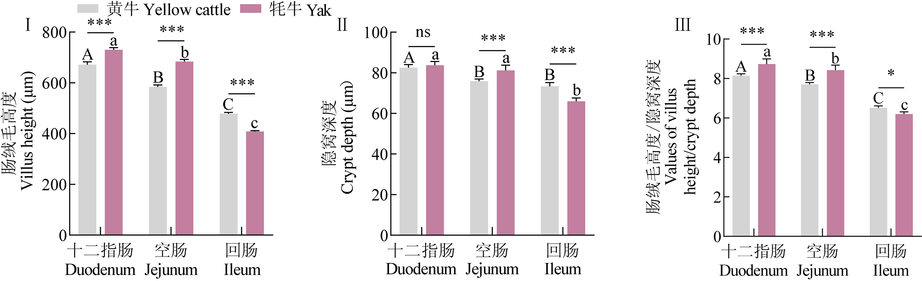
图2
牦牛和黄牛小肠组织形态学测量(H.E染色) (Ⅰ) 肠绒毛高度组织和物种差异对比;(Ⅱ) 隐窝深度组织差异和物种差异对比;(Ⅲ) 消化指标参数V/C组织差异和物种差异对比。其中ns表示物种间无显著差异(P>0.05),*表示物种间差异显著(P<0.05),**表示物种间差异非常显著(P<0.01),***表示物种间差异极其显著(P<0.001);不同大写字母(A、B、C)表示黄牛不同小肠组织间差异显著(P<0.05),不同小写字母(a、b、c)表示牦牛不同小肠组织间差异显著(P<0.05)。下同"

图3
牦牛和黄牛小肠组织切片(PAS染色) A—C、J—L依次为黄牛和牦牛的十二指肠、空肠和回肠的PAS染色, 200×; D—F、M—O依次为黄牛和牦牛的十二指肠、空肠和回肠的PAS染色, 400×; G—I、P—R依次为黄牛和牦牛的十二指肠、空肠和回肠的PAS染色, 1000×"
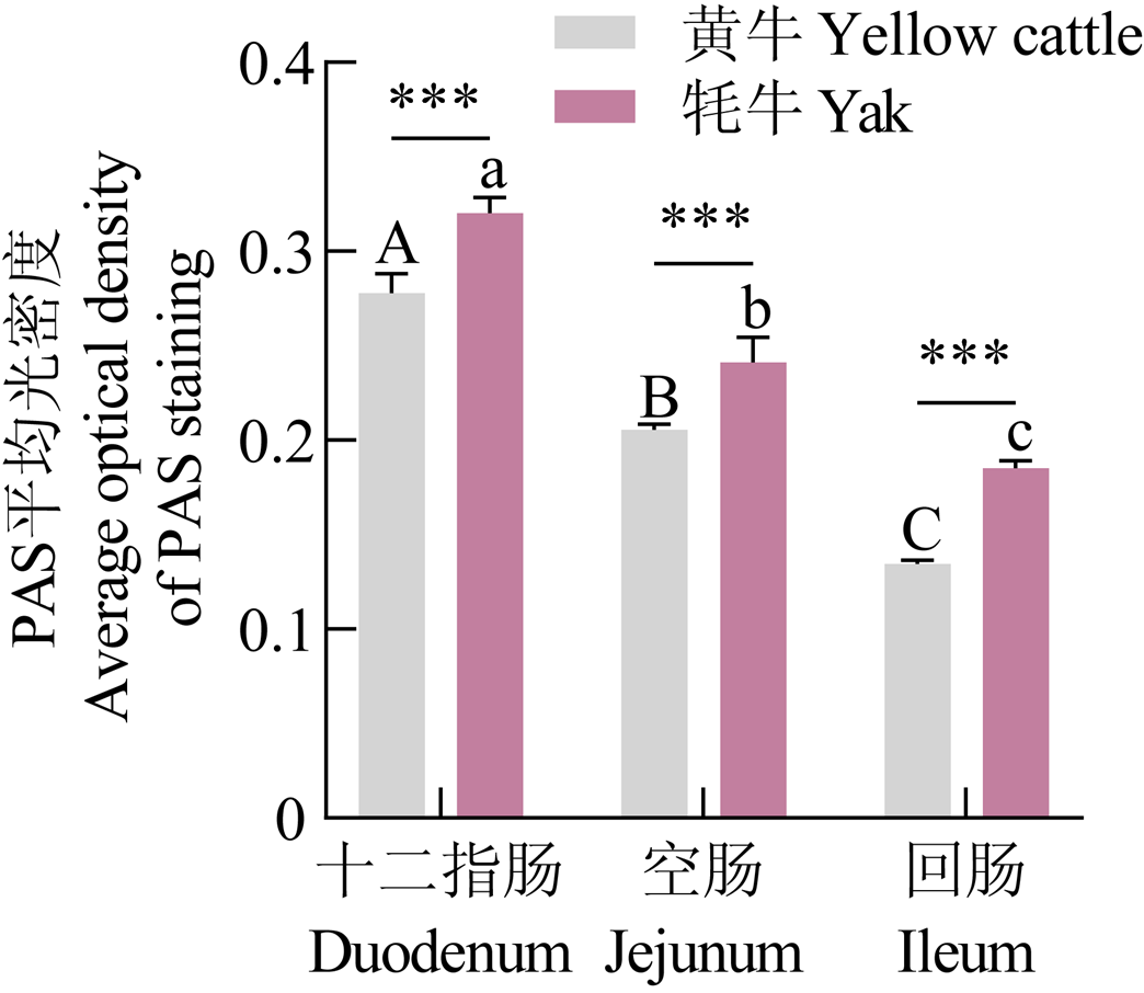
图4
牦牛和黄牛小肠组织形态学测量(PAS染色)"

图5
牦牛和黄牛小肠组织切片(Masson染色) A—C、J—L依次为黄牛和牦牛的十二指肠、空肠和回肠的Masson染色, 200×; D—F、M—O依次为黄牛和牦牛的十二指肠、空肠和回肠的Masson染色, 400×; G—I、P—R依次为黄牛和牦牛的十二指肠、空肠和回肠的Masson染色, 1000×"
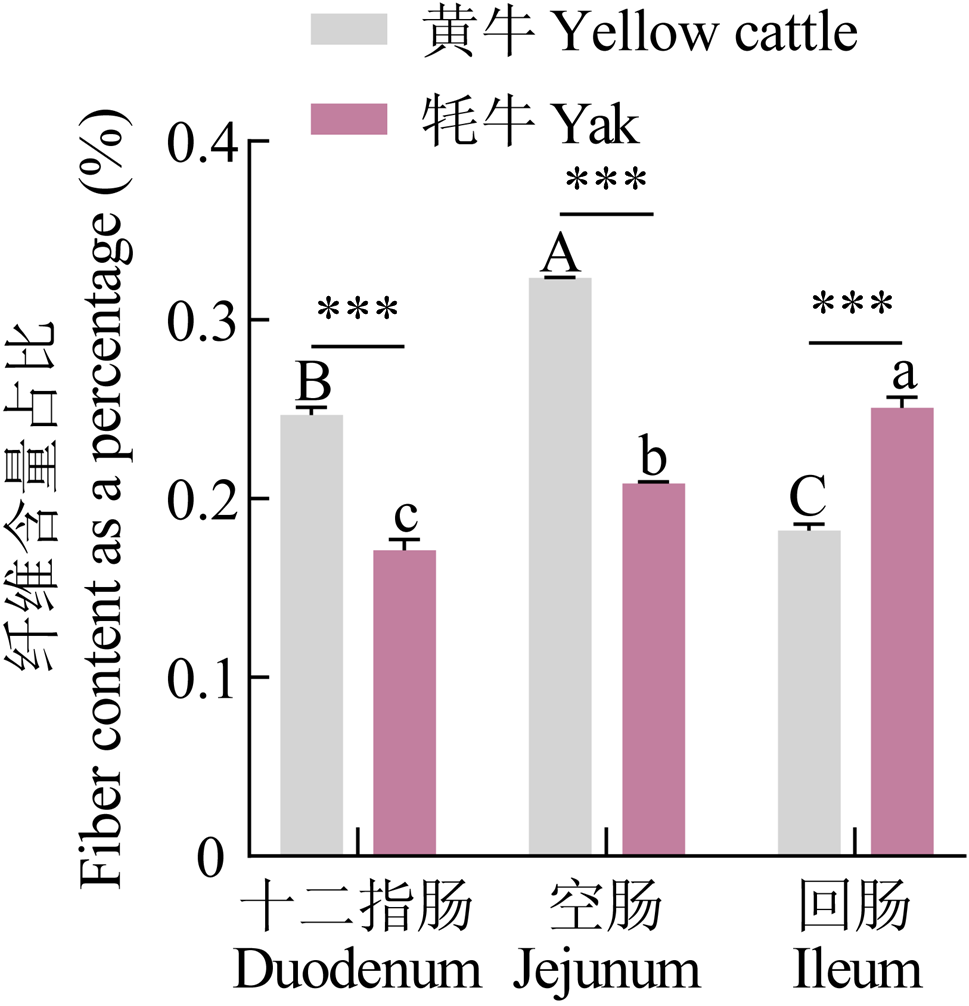
图6
牦牛和黄牛小肠组织形态学测量(Masson染色)"

图7
牦牛和黄牛小肠组织切片(阴性对照) A—C、J—L依次为黄牛和牦牛的十二指肠、空肠和回肠的阴性对照组, 200×; D—F、M—O依次为黄牛和牦牛的十二指肠、空肠和回肠的阴性对照组, 400×; G—I、P—R依次为黄牛和牦牛的十二指肠、空肠和回肠的阴性对照组, 1000×"

图8
牦牛和黄牛小肠组织切片(PDK1染色) A—C、J—L依次为黄牛和牦牛的十二指肠、空肠和回肠的PDK1染色, 200×; D—F、M—O依次为黄牛和牦牛的十二指肠、空肠和回肠的PDK1染色, 400×; G—I、P—R依次为黄牛和牦牛的十二指肠、空肠和回肠的PDK1染色, 1000×"
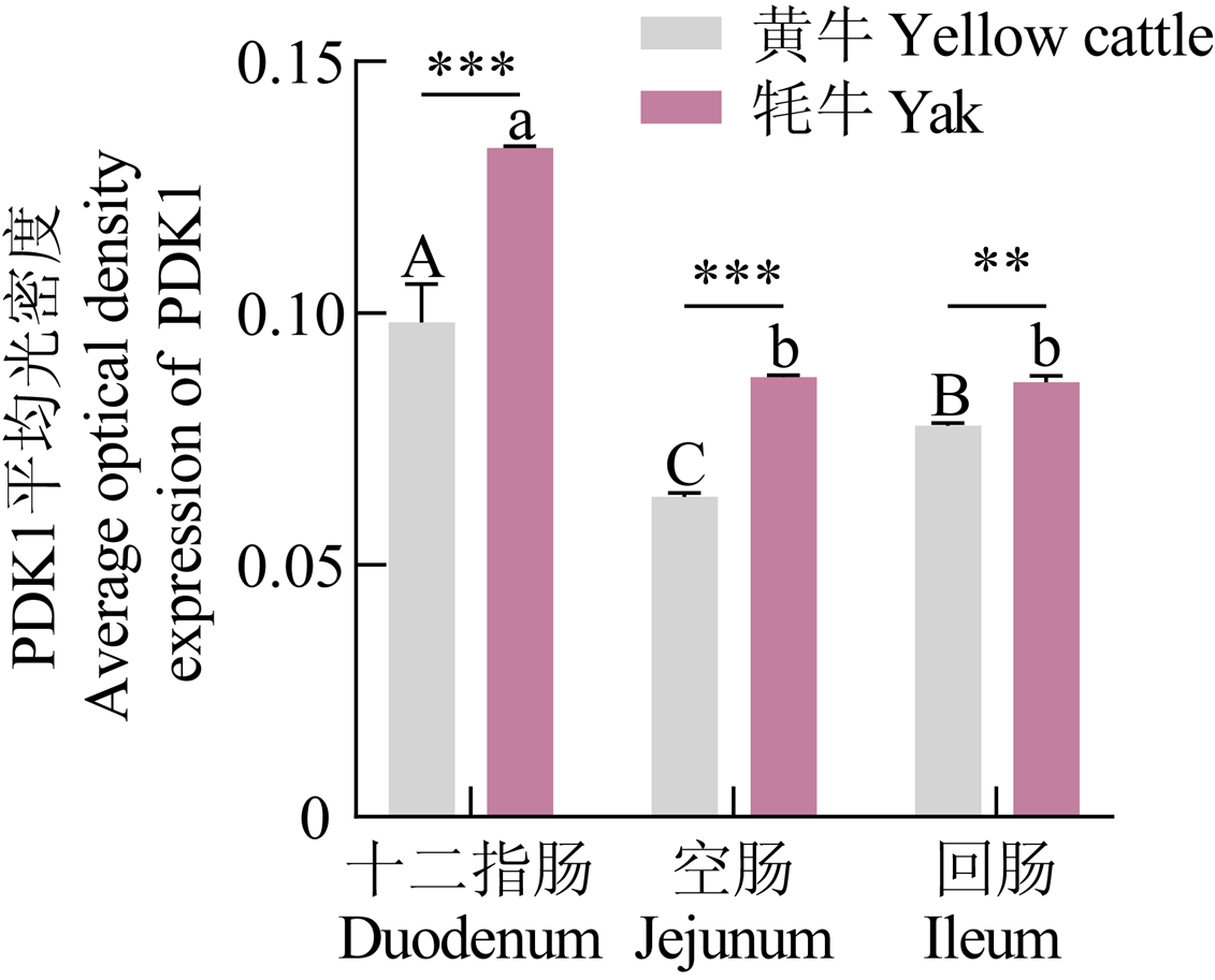
图9
牦牛和黄牛小肠组织形态学测量(PDK1染色)"

图10
牦牛和黄牛小肠组织切片(HIF-1α染色) A—C、J—L依次为黄牛和牦牛的十二指肠、空肠和回肠的HIF-1α染色, 200×; D—F、M—O依次为黄牛和牦牛的十二指肠、空肠和回肠的HIF-1α染色, 400×; G—I、P—R依次为黄牛和牦牛的十二指肠、空肠和回肠的HIF-1α染色, 1000×"
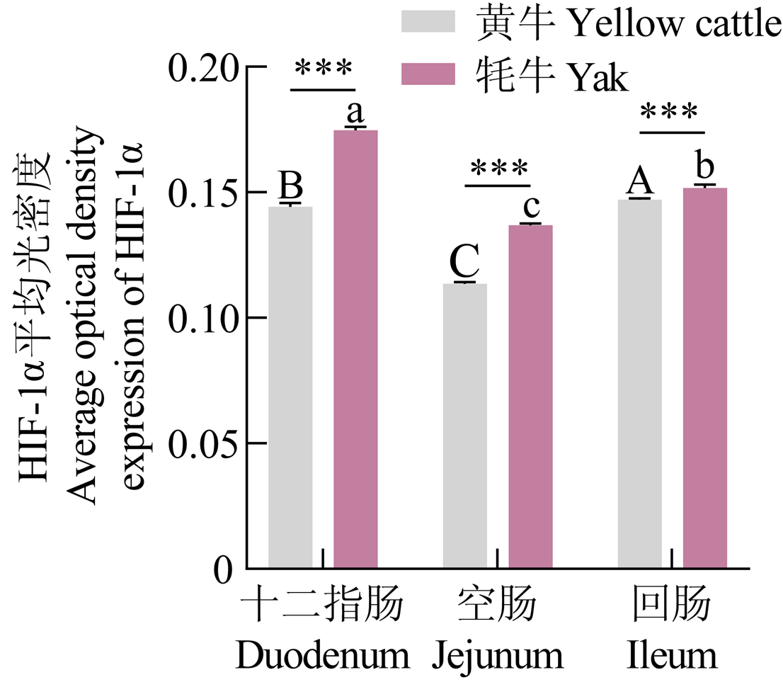
图11
牦牛和黄牛小肠组织形态学测量(HIF-1α染色)"

图12
牦牛和黄牛小肠组织切片(VEGF染色) A—C、J—L依次为黄牛和牦牛的十二指肠、空肠和回肠的VEGF染色, 200×; D—F、M—O依次为黄牛和牦牛的十二指肠、空肠和回肠的VEGF染色, 400×; G—I、P—R依次为黄牛和牦牛的十二指肠、空肠和回肠的VEGF染色, 1000×"
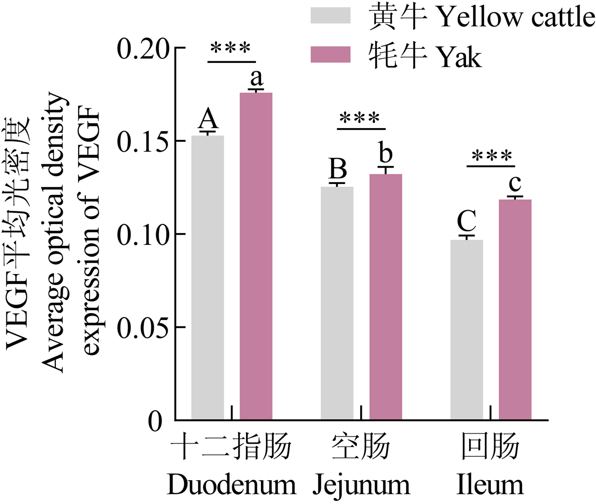
图13
牦牛和黄牛小肠组织形态学测量(VEGF染色)"

图14
牦牛和黄牛小肠组织切片(TGF-β染色) A—C、J—L依次为黄牛和牦牛的十二指肠、空肠和回肠的TGF-β染色, 200×; D—F、M—O依次为黄牛和牦牛的十二指肠、空肠和回肠的TGF-β染色, 400×; G—I、P—R依次为黄牛和牦牛的十二指肠、空肠和回肠的TGF-β染色, 1000×"
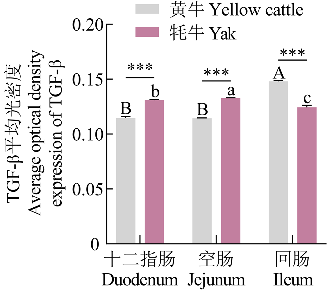
图15
牦牛和黄牛小肠组织形态学测量(TGF-β染色)"

图16
牦牛和黄牛小肠组织切片(Smad2染色) A—C、J—L依次为黄牛和牦牛的十二指肠、空肠和回肠的Smad2染色, 200×; D—F、M—O依次为黄牛和牦牛的十二指肠、空肠和回肠的Smad2染色, 400×; G—I、P—R依次为黄牛和牦牛的十二指肠、空肠和回肠的Smad2染色, 1000×"
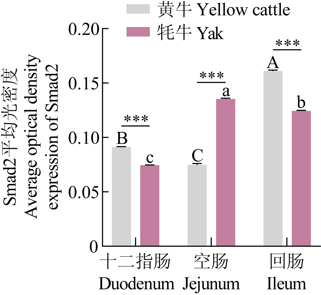
图17
牦牛和黄牛小肠组织形态学测量(Smad2染色)"

图18
牦牛和黄牛小肠组织切片(Smad3染色) A—C、J—L依次为黄牛和牦牛的十二指肠、空肠和回肠的Smad3染色, 200×; D—F、M—O依次为黄牛和牦牛的十二指肠、空肠和回肠的Smad3染色, 400×; G—I、P—R依次为黄牛和牦牛的十二指肠、空肠和回肠的Smad3染色, 1000×"
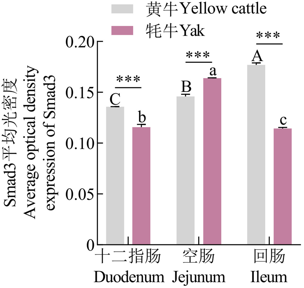
图19
牦牛和黄牛小肠组织形态学测量(Smad3染色)"
| [1] |
杨琦玥, 黄勇, 陈亚冰, 刘洛川, 李键, 兰道亮. 16S rRNA高通量测序技术筛选牦牛瘤胃细菌基因组DNA提取方法及菌群结构[J]. 中国农业科学, 2017, 50(5): 932-941. DOI:10.3864/j.issn.0578-1752.2017.05.016.
|
|
|
|
| [2] |
doi: 10.1038/ng.2343 pmid: 22751099 |
| [3] |
doi: 10.1080/19490976.2018.1502539 pmid: 30136893 |
| [4] |
|
| [5] |
doi: 10.1007/s00109-007-0277-z |
| [6] |
doi: 10.1038/s41390-024-03569-7 |
| [7] |
doi: 10.1056/NEJMra0910283 pmid: 21323543 |
| [8] |
doi: 10.1016/j.bbcan.2023.188971 |
| [9] |
pmid: 11642366 |
| [10] |
阿依木古丽·阿不都热依木, 张晨, 蔡勇, 覃圣, 罗文学, 扎西英派. 藏绵羊肺脏结构特点及HIF-1α和AQP1的表达特性[J]. 中国农业科学, 2023, 56(11): 2202-2211. DOI:10.3864/j.issn.0578-1752.2023.11.013.
|
|
|
|
| [11] |
doi: 10.1016/j.gene.2006.08.004 |
| [12] |
doi: 10.1080/10495398.2014.1002563 pmid: 25927169 |
| [13] |
doi: 10.1016/j.ccr.2004.11.022 pmid: 15652751 |
| [14] |
|
| [15] |
|
| [16] |
doi: 10.1165/rcmb.2016-0186OC pmid: 28915065 |
| [17] |
pmid: 11344109 |
| [18] |
doi: 10.1161/01.res.77.3.638 pmid: 7641334 |
| [19] |
doi: 10.1038/nm0603-669 pmid: 12778165 |
| [20] |
doi: 10.1016/j.cmet.2013.08.001 pmid: 23973331 |
| [21] |
doi: 10.1016/j.tips.2021.04.002 pmid: 33966900 |
| [22] |
林凯, 虞德兵, 解晓东, 于敏莉, 李东锋, 杜文兴. TGF-β/Smad信号通路在Follistatin调节鸭骨骼肌卫星细胞增殖过程中的作用机制[J]. 中国农业科学, 2015, 48(12): 2460-2468. DOI: 10.3864/j.issn.0578-1752.2015.12.019.
|
|
|
|
| [23] |
郭泽媛, 杜张胜, 张雅琦, 陈春路, 马晓燕, 成颖, 王锴, 吕丽华. Smad7介导TGF-β信号通路对绵羊卵泡颗粒细胞增殖的影响[J]. 中国农业科学, 2023, 56(13): 2597-2608. DOI: 10.3864/j.issn.0578-1752.2023.13.013.
|
|
|
|
| [24] |
doi: 10.3390/ijms25084465 |
| [25] |
张瑞莉, 葛铭, 郑世民. 缺氧缺血致脑损伤机理的研究[J]. 东北农业大学学报, 2005, 36(6): 774-777.
|
|
|
|
| [26] |
徐一帆, 周陈晨, 刘希成, 郭维华, 周建. 缺氧相关疾病动物模型研究进展[J]. 四川大学学报(医学版), 2025, 56(2): 331-338.
|
|
|
|
| [27] |
pmid: 8378616 |
| [28] |
巨晓军, 章明, 刘一帆, 姬改革, 单艳菊, 屠云洁, 邹剑敏, 张海涛, 卞良永, 束婧婷. 整合肠道菌群与小分子代谢物分析调控肉鸡饲料转化率的影响因素[J]. 中国农业科学, 2025, 58(6): 1223-1238. DOI: 10.3864/j.issn.0578-1752.2025.06.013.
|
|
|
|
| [29] |
doi: 10.1080/19490976.2024.2405547 |
| [30] |
王文婷, 邬应龙. 氧化魔芋葡甘露聚糖对高脂饮食C57BL/6J小鼠十二指肠形态及肠道菌群的影响[J]. 食品科学, 2013, 34(19): 306-310.
doi: 10.7506/spkx1002-6630-201319063 |
|
doi: 10.1111/jfds.1969.34.issue-4 |
|
| [31] |
李蕊彤, 陈清梅, 徐建芳, 张军民, 司玮, 张铁鹰. 基于猪肠道类器官模型的甘草次酸抗氧化功能及机制研究[J]. 中国农业科学, 2025, 58(22): 4771-4785. DOI: 10.3864/j.issn.0578-1752.2025.22.016.
|
|
|
|
| [32] |
doi: 10.1111/imr.12182 pmid: 24942678 |
| [33] |
doi: 10.1146/annurev-biochem-011520-105053 pmid: 32243763 |
| [34] |
pmid: 9753270 |
| [35] |
doi: 10.3168/jds.2021-21723 pmid: 35691749 |
| [36] |
doi: 10.3748/wjg.v9.i4.804 |
| [37] |
pmid: 3096381 |
| [38] |
doi: 10.1016/j.molcel.2010.09.022 pmid: 20965423 |
| [39] |
doi: 10.1016/j.cmet.2006.02.002 |
| [40] |
doi: 10.1080/19490976.2024.2434685 |
| [41] |
doi: 10.1126/scisignal.aav5183 |
| [42] |
doi: 10.1074/jbc.M115.646984 pmid: 26318453 |
| [43] |
doi: 10.1074/jbc.M604507200 |
| [44] |
doi: 10.1038/nature04433 |
| [45] |
doi: 10.1016/j.molcel.2011.05.004 pmid: 21596309 |
| [46] |
doi: 10.1016/j.redox.2015.09.009 |
| [47] |
doi: 10.1096/fsb2.v38.11 |
| [1] | 格日其木格, 普布占堆, 徐青, 侯玲玲. 低氧对牛肾细胞增殖和线粒体自噬的影响[J]. 中国农业科学, 2026, 59(6): 1333-1347. |
| [2] | 刘卓琳, 刘红云. 基于网络药理学和分子对接探究芹菜素缓解奶牛热应激及低氧应激的潜力与机制[J]. 中国农业科学, 2024, 57(5): 1010-1022. |
| [3] | 孟朝轶, 王运路, 姚一龙, 席广银, 牛家强, 索朗斯珠, 郭敏, 徐业芬. lncRNA-MSTRG.7889.1竞争性结合bta-miR-146a靶向Smad4调控牦牛颗粒细胞的凋亡[J]. 中国农业科学, 2024, 57(4): 797-809. |
| [4] | 潘阳阳, 王靖雷, 王萌, 王立斌, 张倩, 陈睿, 张甜甜, 崔燕, 徐庚全, 樊江峰, 余四九. 牦牛早期胚胎核旁斑点形成及对后续发育的影响[J]. 中国农业科学, 2023, 56(6): 1189-1203. |
| [5] | 李继荣, 刘鑫, 王君, 曹晓钢, 次顿. 牦牛乳制品加工过程中稳定碳、氮同位素分馏效应[J]. 中国农业科学, 2023, 56(10): 1982-1993. |
| [6] | 阿依木古丽·阿不都热依木,阿尔祖古丽·阿依丁,王家敏,石嘉琛,马芳芳,蔡勇,乔自林. 大豆异黄酮对牦牛卵巢颗粒细胞增殖和凋亡的影响[J]. 中国农业科学, 2022, 55(8): 1667-1675. |
| [7] | 冉宏标,赵丽玲,王会,柴志欣,王吉坤,王嘉博,武志娟,钟金城. LncFAM200B对牦牛肌内前体脂肪细胞脂质沉积的影响[J]. 中国农业科学, 2022, 55(13): 2654-2666. |
| [8] | 陈露露,王会,王吉坤,王嘉博,柴志欣,陈智华,钟金城. 藏黄牛与宣汉黄牛心脏miRNA表达谱比较[J]. 中国农业科学, 2020, 53(8): 1677-1687. |
| [9] | 李雪茹,师希雄,王建忠,张攀高,田铸,韩玲. 一氧化氮合成酶抑制剂对宰后成熟过程中牦牛肉品质的影响[J]. 中国农业科学, 2020, 53(8): 1617-1626. |
| [10] | 苗建军,彭忠利,高彦华,柏雪,谢昕廷. 日粮中添加小肽对育肥牦牛生产性能和消化道PepT1 mRNA表达的影响[J]. 中国农业科学, 2020, 53(23): 4950-4960. |
| [11] | 潘阳阳,王萌,芮弦,王立斌,何翃闳,王靖雷,马睿,徐庚全,崔燕,樊江峰,余四九. IGF-1调控RBM3表达抑制低温应激诱导牦牛卵丘细胞凋亡[J]. 中国农业科学, 2020, 53(11): 2285-2296. |
| [12] | 孙凯, 李冬秀, 杨靖, 董骥驰, 严贤诚, 罗立新, 刘永柱, 肖武名, 王慧, 陈志强, 郭涛. 水稻耐淹成苗率相关性状全基因组的关联分析[J]. 中国农业科学, 2019, 52(3): 385-398. |
| [13] | 富丽霞,马涛,刁其玉,成述儒,宋雅喆,孙卓琳. 肉羊精料可代谢蛋白质预测模型的建立[J]. 中国农业科学, 2019, 52(3): 539-549. |
| [14] | 黄向月,熊显荣,韩杰,杨显英,王艳,王斌,李键. KDM1A在牦牛卵泡发育过程中的表达[J]. 中国农业科学, 2019, 52(24): 4624-4631. |
| [15] | 曲星梅,薛复来,黄晓瑜,张宇,邢晓楠,张恩平. 断奶日龄和日粮营养水平对陕北白绒山羊小肠 形态发育和消化酶活性的影响[J]. 中国农业科学, 2019, 52(19): 3460-3470. |
|
||
