| [1] |
LI M X, XIAO N, LI J N, LIN H X, FAN H J. Evaluation of immune efficacy of Omp2 protein against Lawsonia intracellularis in mice. Veterinary Microbiology, 2021, 263: 109274.
|
| [2] |
LAWSON G H K, GEBHART C J. Proliferative enteropathy. Journal of Comparative Pathology, 2000, 122(2/3): 77-100.
|
| [3] |
陈健. 猪增生性肠炎的流行病学、临床症状、鉴别诊断及防控. 现代畜牧科技, 2021(12): 96-97.
|
|
CHEN J. Epidemiology, clinical symptoms, differential diagnosis, prevention and control of porcine proliferative enteropathy. Modern Animal Husbandry Science & Technology, 2021(12): 96-97. (in Chinese)
|
| [4] |
KROLL J J, ROOF M B, HOFFMAN L J, DICKSON J S, HANK HARRIS D L. Proliferative enteropathy: a global enteric disease of pigs caused by Lawsonia intracellularis. Animal Health Research Reviews, 2005, 6(2): 173-197.
|
| [5] |
OBRADOVIC M R, WILSON H L. Immune response and protection against Lawsonia intracellularis infections in pigs. Veterinary Immunology and Immunopathology, 2020, 219: 109959.
|
| [6] |
BIESTER H E, SCHWARTE L H. Intestinal adenoma in swine. American Journal of Pathology, 1931, 7(2): 175-185.6.
pmid: 19969959
|
| [7] |
WON G, CHI N K, PARK Y. The effectiveness of commercial vaccination against Lawsonia intracellularis in mitigating the reduction in ADWG, the increased mortality and fecal shedding of the vaccinated pigs: A systematic review and meta-analysis. Veterinary Sciences, 2022, 9(10): 536.
|
| [8] |
肖宁. 胞内劳森菌弱毒疫苗研制及其胞内存活机制[D]. 南京: 南京农业大学, 2022.
|
|
XIAO N. Development of a live attenuated vaccine against Lawsonia intracellularis and the mechanism of intracellular survival[D]. Nanjing: Nanjing Agricultural University, 2022. (in Chinese)
|
| [9] |
KROLL J J, ROOF M B, MCORIST S. Evaluation of protective immunity in pigs following oral administration of an avirulent live vaccine of Lawsonia intracellularis. American Journal of Veterinary Research, 65(5): 559-565.
|
| [10] |
ROERINK F, MORGAN C L, KNETTER S M, PASSAT M H, ARCHIBALD A L, AIT-ALI T, STRAIT E L. A novel inactivated vaccine against Lawsonia intracellularis induces rapid induction of humoral immunity, reduction of bacterial shedding and provides robust gut barrier function. Vaccine, 2018, 36(11): 1500-1508.
|
| [11] |
XIAO N, LI J N, LI M X, ZHOU H, LIN H X, FAN H J. Isolation and in vitro cultivation of Lawsonia intracellularis from China. Veterinary Microbiology, 2022, 270: 109457.
|
| [12] |
PEDERSEN K S, HOLYOAKE P, STEGE H, NIELSEN J P. Diagnostic performance of different fecal Lawsonia intracellularis: Specific polymerase chain reaction assays as diagnostic tests for proliferative enteropathy in pigs: A review. Journal of Veterinary Diagnostic Investigation, 2010, 22(4): 487-494.
|
| [13] |
|
|
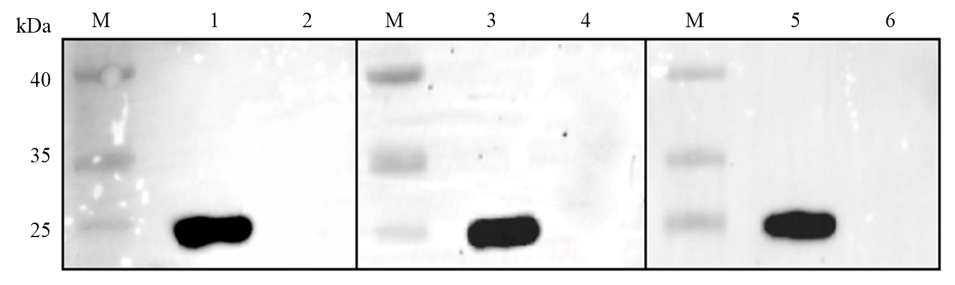
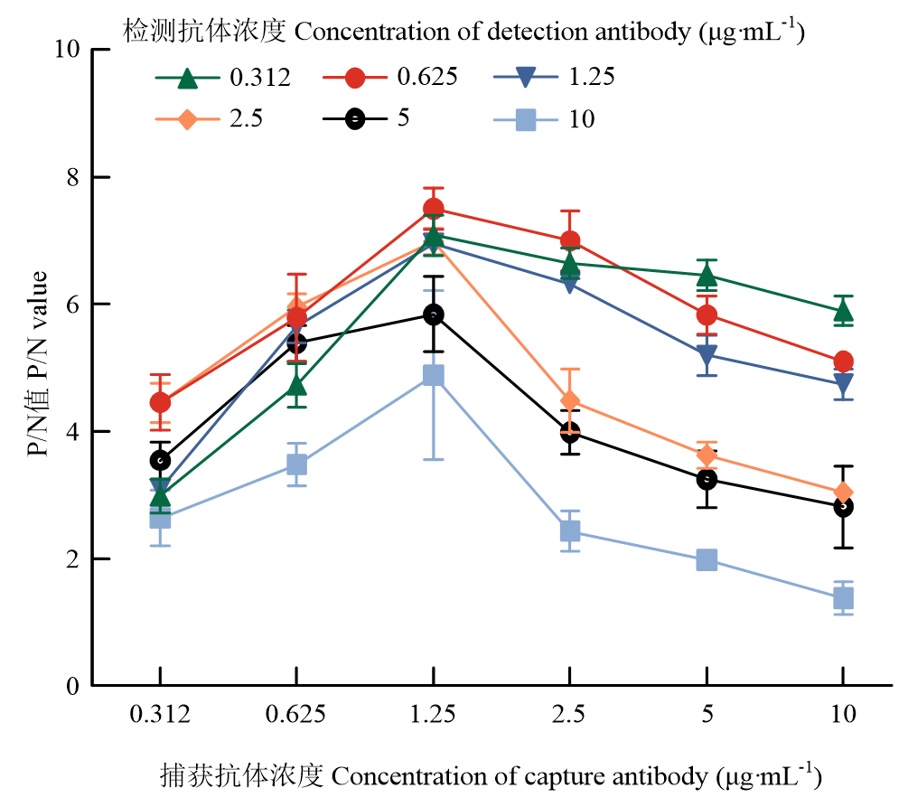
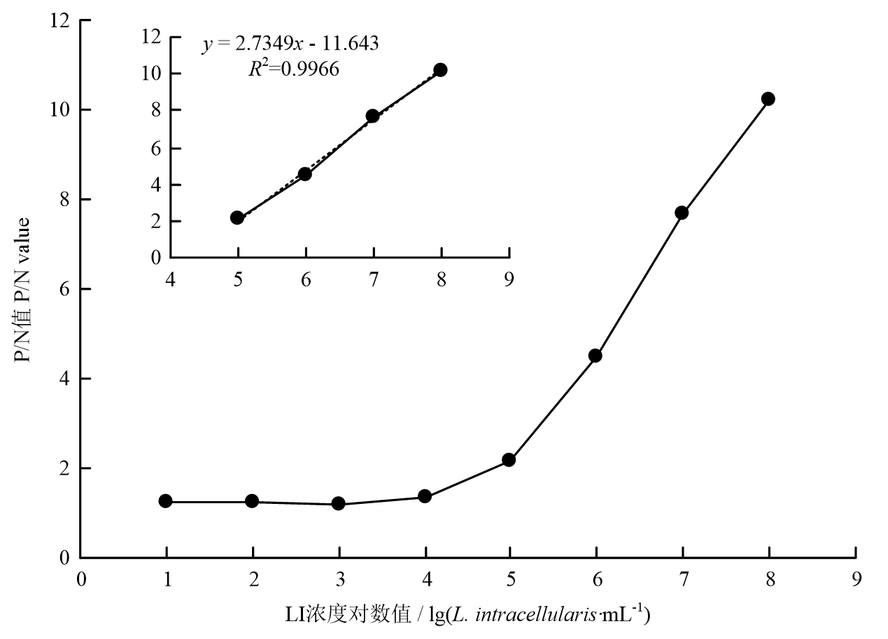
ZHANG C Y, ZHOU H, LIN H X, FAN H J. Development and application of indirect ELISA kits for antibody detection of porcine proliferative enteropathy. Scientia Agricultura Sinica, 2024, 57(16): 3283-3293. doi: 10.3864/j.issn.0578-1752.2024.16.014. (in Chinese)
|
| [14] |
WATTANAPHANSAK S, ASAWAKARN T, GEBHART C J, DEEN J. Development and validation of an enzyme-linked immunosorbent assay for the diagnosis of porcine proliferative enteropathy. Journal of Veterinary Diagnostic Investigation, 2008, 20(2): 170-177.
pmid: 18319429
|
| [15] |
CORZO C A, FRIENDSHIP R, DEWEY C, BLACKWELL T. Comparison of 2 serologic tests for the diagnosis of porcine proliferative enteropathy. La Revue Veterinaire Canadienne, 2005, 46(5): 433-435.
|
| [16] |
|
|
LI M X, LI J N, ZHOU H, XIAO N, LIN H X, MA Z, FAN H J. Establishment and preliminary application of Lawsonia intracellularis IPMA antigen detection method based on sod C monoclonal antibody. Scientia Agricultura Sinica, 2021, 54(20): 4478-4486. doi: 10.3864/j.issn.0578-1752.2021.20.020. (in Chinese)
|
| [17] |
RICHTER B, LADINIG A, NEDOROST N, HERBERT W. A TaqMan quantitative polymerase chain reaction assay for the detection of Lawsonia intracellularis in fecal and tissue samples from pigs. Journal of Veterinary Diagnostic Investigation, 2010, 22(1): 70-73.
|
| [18] |
GUEDES R M, GEBHART C J, WINKELMAN N L, MACKIE- NUSS R A, MARSTELLER T A, DEEN J. Comparison of different methods for diagnosis of porcine proliferative enteropathy. Revue Canadienne de Recherche Veterinaire, 2002, 66(2): 99-107.
|
| [19] |
JACOBSON M, ASPAN A, KÖNIGSSON M H, SEGERSTAD C H A, WALLGREN P, FELLSTRÖM C, JENSEN-WAERN M, GUNNARSON A. Routine diagnostics of Lawsonia intracellularis performed by PCR, serological and post mortem examination, with special emphasis on sample preparation methods for PCR. Veterinary Microbiology, 2004, 102(3/4): 189-201.
|
| [20] |
XIAO N, LI J N, LI M X, HU Y T, LIN H X, FAN H J. Preparation and characterization of a new monoclonal antibody specific against Lawsonia intracellularis and its application in indirect immunofluorescence and immunocytochemistry assay. Frontiers in Veterinary Science, 2021, 8: 753610.
|
| [21] |
庄金秋, 梅建国, 姚春阳, 张颖, 李峰. 猪增生性肠炎实验室诊断方法研究进展. 养猪, 2022(6): 121-128.
|
|
ZHUANG J Q, MEI J G, YAO C Y, ZHANG Y, LI F. Research progress on the diagnosis methods of the porcine proliferative enteritis in laboratory. Swine Production, 2022(6): 121-128. (in Chinese)
|
| [22] |
XU L L, CHEN Z H, GONG H Y, PEI X X, ZHU Y Y, LU Y C, WANG Y M, NAN S F, YIN Y P, ZHAO Q, FAN Y P, SUN Y N, XIAO S Q. Development a high-sensitivity sandwich ELISA for determining antigen content of porcine circovirus type 2 vaccines. Journal of Virological Methods, 2024, 328: 114954.
|
| [23] |
CHOI S Y, RHIE G E, JEON J H. Development of a double-antibody sandwich ELISA for sensitive detection of Yersinia pestis. Microbiology and Immunology, 2020, 64(1): 72-75.
|
| [24] |
田瑞雨, 熊祺琰, 刘茂军, 韦艳娜, 武昱孜, 冯志新, 姜平, 邵国青. 猪肺炎支原体抗原定量ELISA检测方法的建立. 中国兽医科学, 2017, 47(4): 471-475.
|
|
TIAN R Y, XIONG Q Y, LIU M J, WEI Y N, WU Y Z, FENG Z X, JIANG P, SHAO G Q. Development of a quantitative enzyme- linked immunosorbent assay for the detection of Mycoplasma hyopneumoniae. Chinese Veterinary Science, 2017, 47(4): 471-475. (in Chinese)
|
| [25] |
彭忠, 涂志勤, 梁婉, 胡睿铭, 王豪男, 寿琎, 董超, 李宗华, 陈焕春, 吴斌. 胞内劳森菌巢式PCR检测方法的建立及初步临床应用. 中国兽医学报, 2017, 37(1): 40-46.
|
|
PENG Z, TU Z Q, LIANG W, HU R M, WANG H N, SHOU J, DONG C, LI Z H, CHEN H C, WU B. Development and preliminary application of a nested PCR menthod for detection of Lawsonia intracellularis in feces of pigs. Chinese Journal of Veterinary Science, 2017, 37(1): 40-46. (in Chinese)
|
| [26] |
WANG J, ZHANG S F, NI W H, ZHAI X Y, XIE F, YUAN H Y, GAO S J, TAI G X. Development and application of a double- antibody sandwich ELISA kit for the detection of serum MUC1 in lung cancer patients. Cancer Biomarkers, 17(4): 369-376.
|
| [27] |
张海洋, 刘爽, 戚雪芹, 王权, 陈兆国, 韩先干, 蒋蔚. 单核细胞增生李斯特菌的单克隆抗体的制备及双抗体夹心ELISA方法的建立. 中国兽医科学, 2021, 51(8): 962-969.
|
|
ZHANG H Y, LIU S, QI X Q, WANG Q, CHEN Z G, HAN X G, JIANG W. Preparation of monoclonal antibodies against Listeria monocytogenes and establishment of a double-antibody sandwich enzyme-linked immunosorbent assay for detection of L. monocytogenes. Chinese Veterinary Science, 2021, 51(8): 962-969. (in Chinese)
|
| [28] |
ZHANG X, ZHU H P, ZHENG X, JIAO Y J, NING L L, ZHOU E M, MU Y. A double-antibody sandwich ELISA for sensitive and specific detection of swine fibrinogen-like protein 1. Frontiers in Immunology, 2021, 12: 670626.
|
| [29] |
WANG W, LI J Z, FAN B C, ZHANG X H, GUO R L, ZHAO Y X, ZHOU J M, ZHOU J Z, SUN D B, LI B. Development of a novel double antibody sandwich ELISA for quantitative detection of porcine deltacoronavirus antigen. Viruses, 2021, 13(12): 2403.
|
| [30] |
张飞雁, 秦涛, 谢红玲, 朱薇, 李建, 郑佳, 冯钊. 猪肺炎支原体p46蛋白定量检测双抗夹心ELISA方法的建立. 动物医学进展, 2024, 45(11): 64-69.
|
|
ZHANG F Y, QIN T, XIE H L, ZHU W, LI J, ZHENG J, FENG Z. Establishment of double antibody sandwich enzyme-linked immunosorbent assay for quantitative detection of p46 protein of Mycoplasma hyopneumoniae. Progress in Veterinary Medicine, 2024, 45(11): 64-69. (in Chinese)
|
| [31] |
WEI Y N, KHOZA T, YU Y F, WANG L, LIU B B, WANG J, GAN L X, HAO F, SHAO G Q, FENG Z X, XIONG Q Y. Development of an indirect competitive enzyme linked immunosorbent assay for the quantitative detection of Mycoplasma hyopneumoniae during the vaccine production process. Journal of Immunological Methods, 2022, 500: 113196.
|
| [32] |
GUEDES R M C, GEBHART C J. Onset and duration of fecal shedding, cell-mediated and humoral immune responses in pigs after challenge with a pathogenic isolate or attenuated vaccine strain of Lawsonia intracellularis. Veterinary Microbiology, 2003, 91(2/3): 135-145.
|
| [33] |
NOGUEIRA M G, COLLINS A M, DONAHOO M, EMERY D. Immunological responses to vaccination following experimental Lawsonia intracellularis virulent challenge in pigs. Veterinary Microbiology, 2013, 164(1/2): 131-138.
|


 ), 周红, 蔺辉星, 范红结(