


中国农业科学 ›› 2021, Vol. 54 ›› Issue (21): 4664-4676.doi: 10.3864/j.issn.0578-1752.2021.21.015
陈慧芳( ),黄绮亮,胡智超,潘晓婷,吴志胜,白银山()
收稿日期:2020-09-27
接受日期:2021-04-25
出版日期:2021-11-01
发布日期:2021-11-09
联系方式:
联系方式:陈慧芳,E-mail: chenhuifang07@163.com。
基金资助:
CHEN HuiFang(),HUANG QiLiang,HU ZhiChao,PAN XiaoTing,WU ZhiSheng,BAI YinShan()
Received:2020-09-27
Accepted:2021-04-25
Published:2021-11-01
Online:2021-11-09
摘要:
【目的】通过分析成熟卵泡液外泌体(mature follicular fiuid Exosomes, mffEXs)和闭锁卵泡液外泌体(atretic follicular fiuid Exosomes, affEXs)miRNA的表达差异,探索卵泡液外泌体(EXs)miRNA在卵泡发育和闭锁过程中的调控作用。【方法】本研究通过抽提4—6 mm猪成熟发育和闭锁卵泡的卵泡液分离外泌体,进行粒径分析及Western Blot检测对EXs进行鉴定,接下来对特征性EXs携带的miRNA测序和功能富集分析,筛选关键信号通路和差异基因。最后,将mffEXs和affEXs作为添加剂进行颗粒细胞培养,利用Q-PCR检测技术分析关键基因的表达,验证两类卵泡液内EXs miRNA在卵泡发育中的调控功能。【结果】成功分离了mffEXs和affEXs,对比mffEXs测序结果,affEXs中有90个miRNA上调表达,220个miRNA下调表达,表明了卵泡液中的miRNA表达水平与调控卵泡发育有关;KEGG富集分析结果显示两类卵泡的差异信号通路主要集中在Ras、cAMP、P53和MAPK等信号通路,涉及调控卵母细胞发育、减数分裂以及颗粒细胞细胞周期等生物学功能。在闭锁卵泡中,上调表达的ssc-let-7a和ssc-miR-133a-3p分别潜在靶向调控细胞周期蛋白依赖性激酶(CDK1)和胰岛素生长因子(IGF1),抑制了G1和G2/M期的运转和类固醇激素代谢,促使颗粒细胞周期运转受阻和颗粒细胞凋亡,引起卵泡闭锁的发生;下调的ssc-miR-21-5p潜在靶向肿瘤抑癌基因(P53),抑制细胞周期运转,促使颗粒细胞凋亡。在体外培养的颗粒细胞中分别添加mffEXs和affEXs,Q-PCR结果显示CDK1在mffEXs中显著上调表达,而P53显著下调表达,表明了测序分析结果的可靠性。这些结果均显示了affEXs中miRNA表达水平的变化促使颗粒细胞凋亡和细胞周期阻滞,引起卵泡闭锁。【结论】猪affEXs携带miRNA增加了对CDK1、IGF1和P53的表达调控,抑制颗粒细胞细胞周期运转和类固醇激素代谢等信号通路,引起颗粒细胞凋亡,导致卵泡闭锁。
陈慧芳,黄绮亮,胡智超,潘晓婷,吴志胜,白银山. 外泌体microRNA在猪成熟和闭锁卵泡中的表达差异及功能分析[J]. 中国农业科学, 2021, 54(21): 4664-4676.
CHEN HuiFang,HUANG QiLiang,HU ZhiChao,PAN XiaoTing,WU ZhiSheng,BAI YinShan. Expression Differences and Functional Analysis of Exosomes microRNA in Porcine Mature and Atretic Follicles[J]. Scientia Agricultura Sinica, 2021, 54(21): 4664-4676.
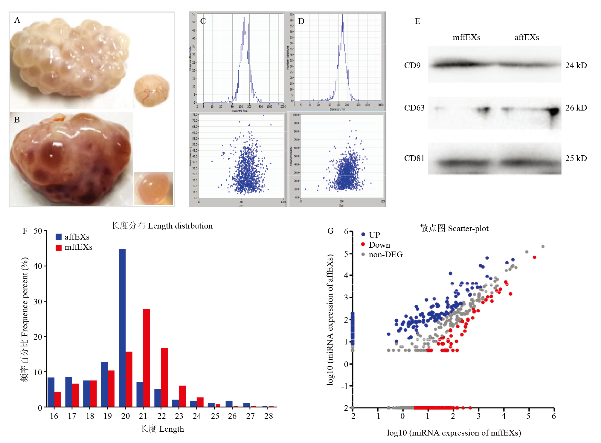
图1
外泌体分离检测与miRNA测序 A:闭锁卵泡的形态观察结果;B:成熟卵泡的形态观察结果;C:mffEXs粒径分析结果;D:affEXs粒径分析结果;E:Westem blot检测结果;F:miRNA的序列长度分布;G:mffEXs和affEXs中miRNA的表达差异结果"
表1
卵泡液EXs中主要差异表达的miRNA"
| miRNA名称 miRNA name | mffEXs-TPM mffEXs-Tags per million | affEXs-TPM affEXs-Tags per million | 差异倍数 Fold change |
|---|---|---|---|
| ssc-miR-1 | 191.1357 | 3683.4041 | 4.268370509 |
| ssc-miR-133a-3p | 125.3327 | 1137.3101 | 3.181790898 |
| ssc-miR-874 | 588.8902 | 4261.314 | 2.855227792 |
| ssc-let-7a | 359.4472 | 2278.7335 | 2.664380439 |
| ssc-let-7f-5p | 378.9345 | 1986.6937 | 2.39034906 |
| ssc-miR-23b | 121.4619 | 631.3819 | 2.378009061 |
| ssc-miR-486 | 810.8585 | 4166.7095 | 2.361386438 |
| ssc-miR-6529 | 548.7143 | 2743.5294 | 2.321905957 |
| ssc-miR-26a | 2160.5543 | 9279.4636 | 2.102639923 |
| ssc-miR-99b | 13585.3175 | 38933.8489 | 1.51897669 |
| ssc-miR-202-3p | 2947.6543 | 7512.8282 | 1.349788776 |
| ssc-miR-1271-5p | 22519.3101 | 52612.4199 | 1.224240778 |
| ssc-miR-21-5p | 555532.4026 | 444467.5 | - 0.321793131 |
| ssc-miR-143 | 1443.9288 | 621.0989 | -1.2171 |
| ssc-miR-10b | 163814.113 | 68106.9853 | -1.266184975 |
| ssc-miR-140-3p | 5264.5074 | 2046.3356 | -1.363255777 |
| ssc-miR-10a-5p | 7077.7606 | 2453.5461 | -1.528424586 |
| ssc-miR-2320-5p | 146.8221 | 49.3588 | -1.572689917 |
| ssc-miR-125b | 13217.7283 | 3973.3874 | -1.734032879 |
| ssc-miR-16 | 371.46 | 109.0008 | -1.768868142 |
| ssc-miR-146a-5p | 6504.7539 | 1748.1259 | -1.895685384 |
| ssc-miR-145-3p | 975.5663 | 257.0773 | -1.924037697 |
| ssc-miR-744 | 367.4557 | 94.6045 | -1.957589617 |
| ssc-miR-184 | 584.4855 | 121.3405 | -2.268106073 |
| ssc-miR-30d | 3433.5021 | 707.4768 | -2.278926102 |
| ssc-miR-132 | 301.9197 | 47.3022 | -2.674185705 |
| ssc-miR-125a | 18571.3967 | 1441.6897 | -3.687249733 |
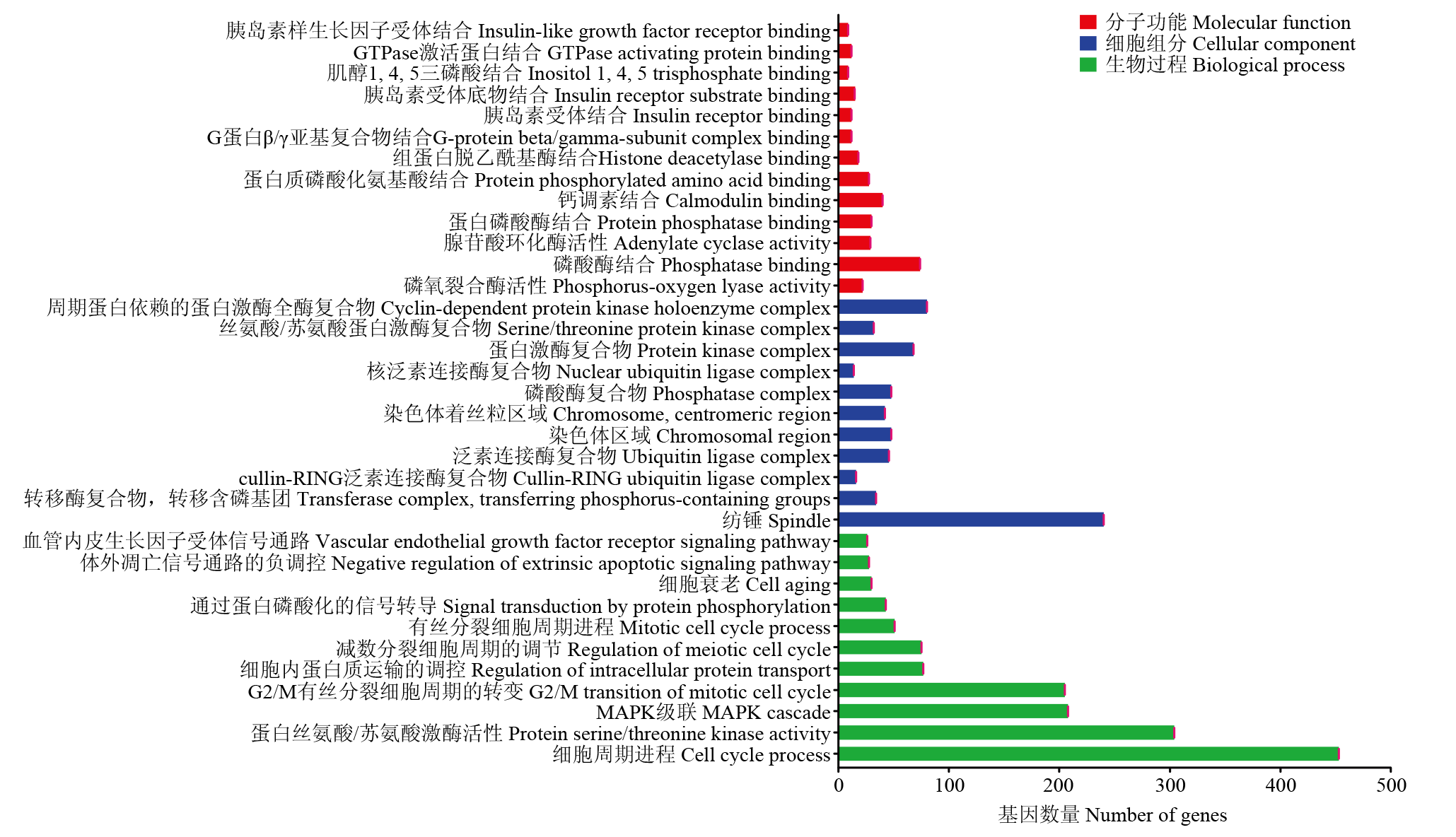
图2
卵泡液EXs中差异miRNA靶基因的GO富集分析"
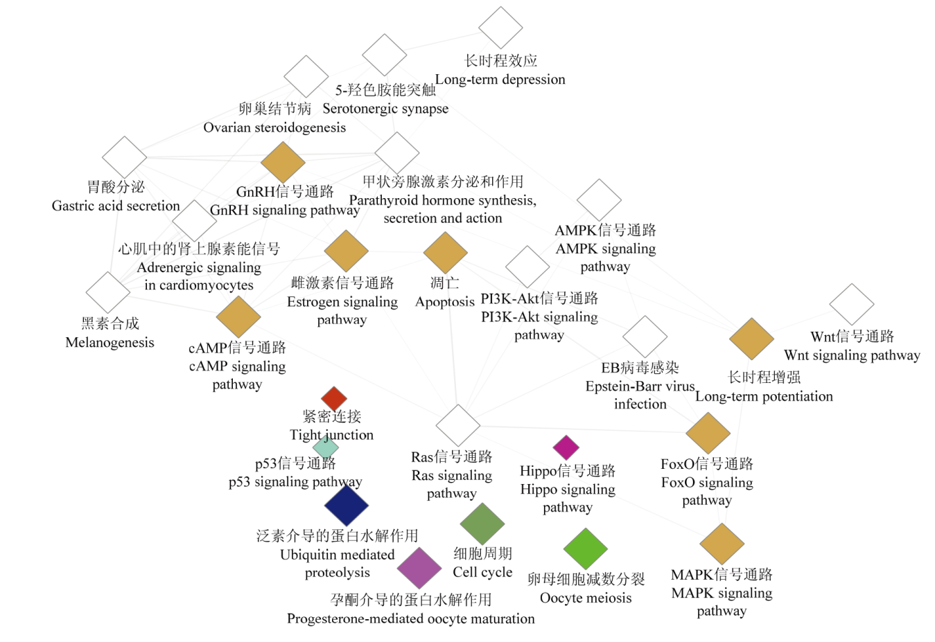
图3
KEGG功能富集分析"
表2
主要信号通路中miRNA调控靶基因信息表"
| 信号通路 Signaling pathways | 靶基因 Target gene list |
|---|---|
| Ras信号通路 Ras signaling pathway (ssc04014) | AKT2 (miR-148b-5p), CALM1 (miR-143-3p), IGF1 (let-7a), INS (miR-424-5p), KRAS (miR-181a), MAP2K1 (miR-1271-5p), MAPK10 (miR-148a-3p), PIK3CB (miR-126-5p), PRKACB (miR-146b), RAF1 (miR-125b) |
| cAMP信号通路 cAMP signaling pathway (ssc04024) | ADCY7 (miR-125b), AKT2 (miR-148b-5p), BRAF (let-7a), CALM1 (miR-130a), CAMK2A (miR-10a-5p), CAMK2G (miR-371-5p), GNAI2 (miR-124a), MAPK10 (miR-1), PDE3B (miR-126-5p), PIK3CG (miR-30a-3p), PPP1CC (miR-1343), PRKACB (miR-181a), RAF1 (miR-424-5) |
| P53信号通路 P53 signaling pathway (ssc04115) | CCNE2 (miR-1), IGF1 (miR-133a-3p), CDK1 (let-7a), CCNB (miR-9791-3p), P53 (miR-21), P53I3 (ssc-miR-7-5p) |
| MAPK信号通路 MAPK signaling pathway (ssc04010) | AKT2 (miR-10a-3p), BRAF (miR-18a), CDC25B (miR-146a-3p), KRAS (miR-181d-5p), MAPK10 (miR-199b-3p), MAPK8 (miR-199a-3p), PPP3CB (miR-202-3p), PRKACB (miR-143-5p), RAF1 (miR-424-5p), RPS6KA3 (miR-125a), P53 (miR-21) |
| 卵母细胞减数分裂 Oocyte meiosis (ssc04114) | ADCY7 (miR-125b), ANAPC1 (miR-326), AR (miR-124a), AURKA (miR-125a), BTRC (miR-1271-5p), CALM1 (miR-181b), CAMK2G (miR-371-5p), CCNE2 (miR-140-3p), CDC26 (miR-218), CDK1 (miR-143-3p), CPEB3 (miR-199b-3p), ESPL1 (miR-141), FBXO11 (miR-129a-3p), IGF1 (miR-133a-3p), ITPR1 (miR-200b), MAP2K1 (miR-143-5p), MAPK12 (miR-125b), MOS (miR-155-3p), PGR (miR-101), PKMYT1 (miR-106a) PLCZ (miR-124a), PPP3CD (miR-199a), PRKACB (miR-143-5p), PTTG1 (miR-1224), RBX (miR-218-3p), REC8 (miR-199b-3p), RPS6KA3 (let-7a), SGOL1 (miR-126-5p), SKP1 (miR-222), SMC1A (miR-128), SMC 3 (miR-9820-5P), STAG3 (miR-27b-3p), YWHAZ (miR-1) |
| 泛素介导的蛋白水解作用 Ubiquitin mediated proteolysis (ssc04120) | IGF1 (miR-133a-3p), BTRC (miR-1), FZR1 (miR-874), CDC27 (miR-1271-5), FBXW11 (miR-671-5p), RBX1 (miR-1224), SKP1 (miR-148a-3p) |
| 细胞周期 Cell cycle (ssc04110) | IGF1 (miR-133a-3p), BUB1 (miR-338), CCNE (miR-140-3p), CDC26 (miR-143-5p), CDK1 (let-7a), FZR1 (miR-138), MAD1L1 (miR-199b-5p), PKMYK1 (miR-106a), PLK1 (miR-18a), SMC1A (miR-132), SMC (miR-9820-5p), YWHAZ (miR-1), P53 (miR-21) |
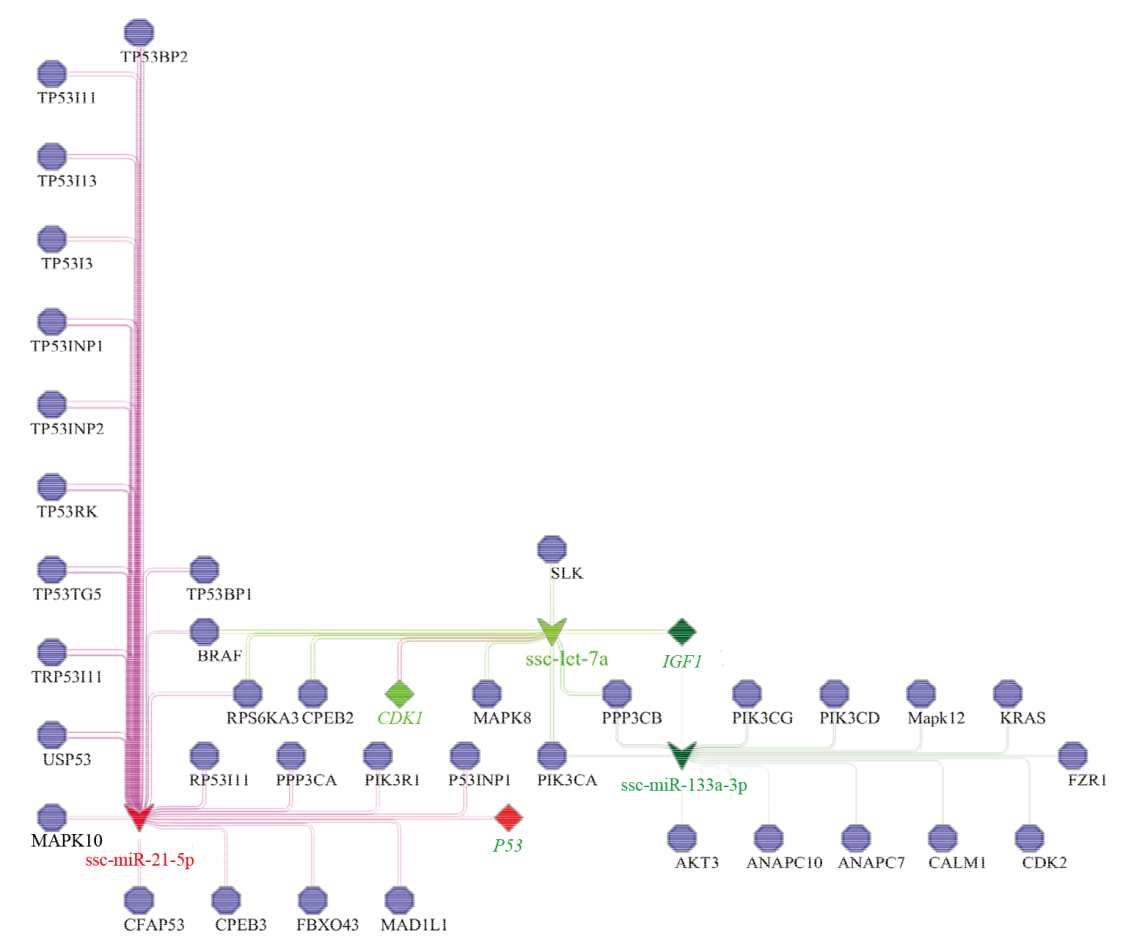
图4
差异性表达miRNA的靶基因互作网络"
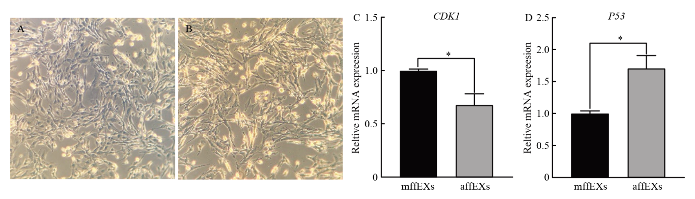
图5
mffEXs和affEXs对猪颗粒细胞调控作用 A:添加mffEXs培养的颗粒细胞结果;B:添加affEXs培养颗粒细胞的结果;C和D:Q-PCR检测CDK1和P53表达结果"
| [1] | JONG E D. Weaning practices and culling policy: Critical steps for optimal reproductive performance of female breeding pigs. 2014. |
| [2] |
FORTUNE J E. Ovarian follicular growth and development in mammals. Biology of Reproduction, 1994, 50(2):225-232.
doi: 10.1095/biolreprod50.2.225 |
| [3] |
DISKIN M G, MACKEY D R, ROCHE J F, SREENAN J M. Effects of nutrition and metabolic status on circulating hormones and ovarian follicle development in cattle. Animal Reproduction Science, 2003, 78(3/4):345-370. doi: 10.1016/s0378-4320(03)00099-x.
doi: 10.1016/s0378-4320(03)00099-x |
| [4] |
HSUEH A J, BILLIG H, TSAFRIRI A. Ovarian follicle atresia: a hormonally controlled apoptotic process. Cancer Management and Research, 1994, 15(6):707-724. doi: 10.1210/edrv-15-6-707.
doi: 10.1210/edrv-15-6-707 |
| [5] |
TILLY J L, TILLY K I. Inhibitors of oxidative stress mimic the ability of follicle-stimulating hormone to suppress apoptosis in cultured rat ovarian follicles. Microorganisms, 1995, 136(1):242-252. doi: 10.1210/endo.136.1.7828537.
doi: 10.1210/endo.136.1.7828537 |
| [6] |
PERSANI L, ROSSETTI R, CACCIATORE C, FABRE S. Genetic defects of ovarian TGF-β-like factors and premature ovarian failure. Journal of Endocrinological Investigation, 2011, 34(3):244-251. doi: 10.1007/BF03347073.
doi: 10.1007/BF03347073 |
| [7] |
SOHEL M M, HOELKER M, NOFERESTI S S, SALILEW- WONDIM D, THOLEN E, LOOFT C, RINGS F, UDDIN M J, SPENCER T E, SCHELLANDER K, TESFAYE D. Exosomal and non-exosomal transport of extra-cellular microRNAs in follicular fluid: Implications for bovine oocyte developmental competence. PLoS ONE, 2013, 8(11):e78505. doi: 10.1371/journal.pone.0078505.
doi: 10.1371/journal.pone.0078505 |
| [8] |
LUO F, JIA R, YING S, WANG Z, WANG F. Analysis of genes that influence sheep follicular development by different nutrition levels during the luteal phase using expression profiling. Animal Genetics, 2016, 47(3):354-364. doi: 10.1111/age.12427.
doi: 10.1111/age.12427 |
| [9] | 胡军和, 唐涛, 谭显胜, 曾智, 吴娟. 外泌体调控卵泡中母细胞发育的研究进展. 中国农学通报, 2019(27):153-157. |
| HU J H, TANG T, TAN X S, ZENG Z, WU J. The advance of exosomes regulating oocyte development in ovarian follicle. Chinese Agricultural Science Bulletin, 2019(27):153-157. (in Chinese) | |
| [10] |
THÉRY C, OSTROWSKI M, SEGURA E. Membrane vesicles as conveyors of immune responses. Nature Reviews Immunology, 2009, 9(8):581-593. doi: 10.1038/nri2567.
doi: 10.1038/nri2567 |
| [11] |
VLASSOV A V, MAGDALENO S, SETTERQUIST R, CONRAD R. Exosomes: current knowledge of their composition, biological functions, and diagnostic and therapeutic potentials. Biochimica et Biophysica Acta, 2012, 1820(7):940-948. doi: 10.1016/j.bbagen.2012. 03.017.
doi: 10.1016/j.bbagen.2012. 03.017 |
| [12] |
KELLER S, RUPP C, STOECK A, RUNZ S, FOGEL M, LUGERT S, HAGER H D, ABDEL B MS, GUTWEIN P, ALTEVOGT P. CD24 is a marker of exosomes secreted into urine and amniotic fluid. Kidney International, 2007, 72(9):1095-1102.
doi: 10.1038/sj.ki.5002486 |
| [13] |
DE LA TORRE GOMEZ C, GOREHAM R V, BECH SERRA J J, NANN T, KUSSMANN M. “exosomics”-A review of biophysics, biology and biochemistry of exosomes with a focus on human breast milk. Frontiers in Genetics, 2018, 9:92. doi: 10.3389/fgene.2018.00092.
doi: 10.3389/fgene.2018.00092 |
| [14] |
BORIACHEK K, UMER M, ISLAM M N, GOPALAN V, LAM A K, NGUYEN N T, SHIDDIKY M J A. An amplification-free electrochemical detection of exosomal miRNA-21 in serum samples. The Analyst, 2018, 143(7):1662-1669. doi: 10.1039/c7an01843f.
doi: 10.1039/c7an01843f |
| [15] |
TAO W, SUN L, SHI H, CHENG Y, JIANG D, FU B, CONTE M A, GAMMERDINGER W J, KOCHER T D, WANG D. Integrated analysis of miRNA and mRNA expression profiles in tilapia gonads at an early stage of sex differentiation. BMC Genomics, 2016, 17(1):328.
doi: 10.1186/s12864-016-2636-z |
| [16] |
SANG Q, YAO Z, WANG H, FENG R, WANG H, ZHAO X, XING Q, JIN L, HE L, WU L, WANG L. Identification of microRNAs in human follicular fluid: characterization of microRNAs that govern steroidogenesis in vitro and are associated with polycystic ovary syndrome in vivo. The Journal of Clinical Endocrinology and Metabolism, 2013, 98(7):3068-3079. doi: 10.1210/jc.2013-1715.
doi: 10.1210/jc.2013-1715 |
| [17] |
WILLIS G R, CONNOLLY K, LADELL K, DAVIES T S, GUSCHINA I A, RAMJI D, MINERS K, PRICE D A, CLAYTON A, JAMES P E, REES D A. Young women with polycystic ovary syndrome have raised levels of circulating annexin v-positive platelet microparticles. Human Reproduction, 2014, 29(12):2756-2763.
doi: 10.1093/humrep/deu281 |
| [18] |
SØRENSEN A E, WISSING M L, ENGLUND A L M, DALGAARD L T. MicroRNA species in follicular fluid associating with polycystic ovary syndrome and related intermediary phenotypes. The Journal of Clinical Endocrinology & Metabolism, 2016, 101(4):1579-1589.
doi: 10.1210/jc.2015-3588 |
| [19] |
杨倩, 刘兰心, 黄荷凤. 多囊卵巢综合征患者卵泡液外泌体的提取鉴定及其miRNAs的提取和检测. 上海交通大学学报(医学版), 2017, 37(8):1085-1089. doi: 10.3969/j.issn.1674-8115.2017.08.007.
doi: 10.3969/j.issn.1674-8115.2017.08.007 |
|
YANG Q, LIU L X, HUANG H F. Extraction and identification of exosomes in follicular fluid from patients with polycystic ovary syndrome and isolation and detection of miRNAs in exosomes. Journal of Shanghai Jiao Tong University (Medical Science), 2017, 37(8):1085-1089. doi: 10.3969/j.issn.1674-8115.2017.08.007. (in Chinese)
doi: 10.3969/j.issn.1674-8115.2017.08.007 |
|
| [20] |
COTICCHIO G, DAL CANTO M, MIGNINI RENZINI M, GUGLIELMO M C, BRAMBILLASCA F, TURCHI D, NOVARA P V, FADINI R. Oocyte maturation: gamete-somatic cells interactions, meiotic resumption, cytoskeletal dynamics and cytoplasmic reorganization. Human Reproduction Update, 2015, 21(4):427-454. doi: 10.1093/humupd/dmv011.
doi: 10.1093/humupd/dmv011 |
| [21] | 刘凯鲁, 胡梦婷, 蔡令波, 李涵, 杨玮杰, 刘嘉茵, 崔毓桂, 千日成. 多囊卵巢综合征患者卵泡液中6种miRNAs表达的检测. 国际生殖健康/计划生育杂志, 2018, 37(1):5-10. doi: 10.3969/j.issn.1674-1889.2018.01.001. |
| LIU K L, HU M T, CAI L B, LI H, YANG W J, LIU J Y, CUI Y G, QIAN R C. Expressions of six MiRNAs in follicular fluid of patients with polycystic ovary syndrome. International Journal of Reproductive Health/ Family Planning, 2018, 37(1):5-10. doi: 10.3969/j.issn.1674-1889.2018.01.001. (in Chinese) | |
| [22] |
詹小舒, 罗惠娜, 罗冬章, 陈胜锋, 王丙云, 白银山, 陈志胜, 刘璨颖, 计慧琴. 犬脐带间充质干细胞来源外泌体对血管内皮细胞增殖、迁移和凋亡的调控作用. 中国组织工程研究, 2019, 23(29):4637-4643. doi: 10.3969/j.issn.2095-4344.1808.
doi: 10.3969/j.issn.2095-4344.1808 |
|
ZHAN X S, LUO H N, LUO D Z, CHEN S F, WANG B Y, BAI Y S, CHEN Z S, LIU C Y, JI H Q. Effects of exosomes derived from canine umbilical cord mesenchymal stem cells on proliferation, migration and apoptosis of vascular endothelial cells. Journal of Clinical Rehabilitative Tissue Engineering Research, 2019, 23(29):4637-4643. doi: 10.3969/j.issn.2095-4344.1808. (in Chinese)
doi: 10.3969/j.issn.2095-4344.1808 |
|
| [23] |
ZHANG H, XU S, LIU X. microRNA profiling of plasma exosomes from patients with ovarian cancer using high-throughput sequencing. Oncology Letters, 2019, 17(6):5601-5607. doi: 10.3892/ol.2019.10220.
doi: 10.3892/ol.2019.10220 |
| [24] |
WAGNER G P, KIN K, LYNCH V J. Measurement of mRNA abundance using RNA-seq data: RPKM measure is inconsistent among samples. Theory in Biosciences, 2012, 131(4):281-285. doi: 10.1007/s12064-012-0162-3.
doi: 10.1007/s12064-012-0162-3 |
| [25] |
YOUNG M D, WAKEFIELD M J, SMYTH G K, OSHLACK A. Gene ontology analysis for RNA-seq: Accounting for selection bias. Proceedings Biological Sciences, 2010, 11(2):R14. doi: 10.1186/gb- 2010-11-2-r14.
doi: 10.1186/gb- 2010-11-2-r14 |
| [26] |
WANG C L, FAN Y C, CHUN-HSIEN TSENG, CHIU C H, TSAI H J, CHOU C H. Salmonella Enteritidis infection slows steroidogenesis and impedes cell growth in hen granulosa cells. Avian Diseases, 2014, 58(4):511-517. doi: 10.1637/10846-041414-reg.1.
doi: 10.1637/10846-041414-reg.1 |
| [27] |
DA SILVEIRA J, ANDRADE G M, PERECIN F, MEIRELES F V, WINGER Q A, BOUMA G J. Isolation and analysis of exosomal microRNAs from ovarian follicular fluid. Methods in Molecular Biology (Clifton, N J), 2018, 1733:53-63. doi: 10.1007/978-1-4939-7601-0_4.
doi: 10.1007/978-1-4939-7601-0_4 |
| [28] |
HU J, TANG T, ZENG Z, WU J, TAN X S, YAN J. The expression of small RNAs in exosomes of follicular fluid altered in human polycystic ovarian syndrome. PeerJ, 2020, 8:e8640.
doi: 10.7717/peerj.8640 |
| [29] |
VICTOR N T, YONATHAN G, GIOVANNI C, MARIA F B. Extracellular vesicles: new players in lymphomas. International Journal of Molecular Sciences, 2018, 20(1). doi: 10.3390/ ijms20010041.
doi: 10.3390/ ijms20010041 |
| [30] |
LÖTVALL J, HILL A F, HOCHBERG F, BUZÁS E I, VIZIO D D, GARDINER C, GHO Y S, KUROCHKIN I V, MATHIVANAN S, QUESENBERRY P. Minimal experimental requirements for definition of extracellular vesicles and their functions: A position statement from the International Society for Extracellular Vesicles. Journal of Extracellular Vesicles, 2014, 3(1):26913.
doi: 10.3402/jev.v3.26913 |
| [31] |
HUNG W T, NAVAKANITWORAKUL R, KHAN T, ZHANG P, DAVIS J S, MCGINNIS L K, CHRISTENSON L K. Stage-specific follicular extracellular vesicle uptake and regulation of bovine granulosa cell proliferation. Biology of Reproduction, 2017, 97(4):644-655. doi: 10.1093/biolre/iox106.
doi: 10.1093/biolre/iox106 |
| [32] |
HUNG W T, HONG X, CHRISTENSON L K, MCGINNIS L K. Extracellular vesicles from bovine follicular fluid support cumulus expansion. Biology of Reproduction, 2015, 93(5):117. doi: 10.1095/ biolreprod.115.132977.
doi: 10.1095/ biolreprod.115.132977 |
| [33] |
AL-DOSSARY A A, STREHLER E E, MARTIN-DELEON P A. Expression and secretion of plasma membrane Ca2+-ATPase 4a (PMCA4a) during murine Estrus: Association with oviductal exosomes and uptake in sperm. PLoS ONE, 2013, 8(11):e80181.
doi: 10.1371/journal.pone.0080181 |
| [34] |
FIELD S L, DASGUPTA T, CUMMINGS M, ORSI N M. Cytokines in ovarian folliculogenesis, oocyte maturation and luteinisation. Molecular Reproduction and Development, 2014, 81(4):284-314. doi: 10.1002/mrd.22285.
doi: 10.1002/mrd.22285 |
| [35] |
BENTWICH I, AVNIEL A, KAROV Y, AHARONOV R, GILAD S, BARAD O, BARZILAI A, EINAT P, EINAV U, MEIRI E, SHARON E, SPECTOR Y, BENTWICH Z. Identification of hundreds of conserved and nonconserved human microRNAs. Nature Genetics, 2005, 37(7):766-770. doi: 10.1038/ng1590.
doi: 10.1038/ng1590 |
| [36] |
KROL J, LOEDIGE I, FILIPOWICZ W. The widespread regulation of microRNA biogenesis, function and decay. Nature Reviews Genetics, 2010, 11(9):597-610. doi: 10.1038/nrg2843.
doi: 10.1038/nrg2843 |
| [37] |
MASOUMI-DEHGHI S, BABASHAH S, SADEGHIZADEH M. microRNA-141-3p-containing small extracellular vesicles derived from epithelial ovarian cancer cells promote endothelial cell angiogenesis through activating the JAK/STAT3 and NF-κB signaling pathways. Journal of Cell Communication and Signaling, 2020, 14(2):233-244. doi: 10.1007/s12079-020-00548-5.
doi: 10.1007/s12079-020-00548-5 |
| [38] |
MACHTINGER R, RODOSTHENOUS R S, ADIR M, MANSOUR A, RACOWSKY C, BACCARELLI A A, HAUSER R. Extracellular microRNAs in follicular fluid and their potential association with oocyte fertilization and embryo quality: An exploratory study. Journal of Assisted Reproduction and Genetics, 2017, 34(4):525-533. doi: 10.1007/s10815-017-0876-8.
doi: 10.1007/s10815-017-0876-8 |
| [39] |
LIANG M, YAO G, YIN M, LÜ M, TIAN H, LIU L, LIAN J, HUANG X, SUN F. Transcriptional cooperation between p53 and NF-κB p65 regulates microRNA-224 transcription in mouse ovarian granulosa cells. Molecular and Cellular Endocrinology, 2013, 370(1/2):119-129. doi: 10.1016/j.mce.2013.02.014.
doi: 10.1016/j.mce.2013.02.014 |
| [40] |
DA SILVEIRA J C, VEERAMACHANENI D N, WINGER Q A, CARNEVALE E M, BOUMA G J. Cell-secreted vesicles in equine ovarian follicular fluid contain miRNAs and proteins: A possible new form of cell communication within the ovarian follicle. Biology of Reproduction, 2012, 86(3):71. doi: 10.1095/biolreprod.111.093252.
doi: 10.1095/biolreprod.111.093252 |
| [41] |
PIETRO C D. Exosome-mediated communication in the ovarian follicle. Journal of Assisted Reproduction and Genetics, 2016, 33(3):303-311.
doi: 10.1007/s10815-016-0657-9 |
| [42] |
KRYSKO D V, DIEZ-FRAILE A, CRIEL G, SVISTUNOV A A, VANDENABEELE P, D’HERDE K. Life and death of female gametes during oogenesis and folliculogenesis. Apoptosis, 2008, 13(9):1065-1087. doi: 10.1007/s10495-008-0238-1.
doi: 10.1007/s10495-008-0238-1 |
| [43] |
TILLY J L, KOWALSKI K I, JOHNSON A L, HSUEH A J. Involvement of apoptosis in ovarian follicular atresia and postovulatory regression. Thrombosis and Haemostasis, 1991, 129(5):2799-2801. doi: 10.1210/endo-129-5-2799.
doi: 10.1210/endo-129-5-2799 |
| [44] |
LIN F, LI R, PAN Z X, ZHOU B, YU D B, WANG X G, MA X S, HAN J, SHEN M, LIU H L. miR-26b promotes granulosa cell apoptosis by targeting ATM during follicular atresia in porcine ovary. PLoS ONE, 2012, 7(6):e38640. doi: 10.1371/journal.pone.0038640.
doi: 10.1371/journal.pone.0038640 |
| [45] |
SALILEW-WONDIM D, AHMAD I, GEBREMEDHN S, SAHADEVAN S, HOSSAIN M D, RINGS F, HOELKER M, THOLEN E, NEUHOFF C, LOOFT C, SCHELLANDER K, TESFAYE D. The expression pattern of microRNAs in granulosa cells of subordinate and dominant follicles during the early luteal phase of the bovine estrous cycle. PLoS ONE, 2014, 9(9):e106795. doi: 10.1371/journal. pone.0106795.
doi: 10.1371/journal. pone.0106795 |
| [46] |
SONTAKKE S D, MOHAMMED B T, MCNEILLY A S, DONADEU F X. Characterization of microRNAs differentially expressed during bovine follicle development. Reproduction (Cambridge, England), 2014, 148(3):271-283. doi: 10.1530/rep-14-0140.
doi: 10.1530/rep-14-0140 |
| [47] |
CARLETTI M Z, FIEDLER S D, CHRISTENSON L K. microRNA 21 blocks apoptosis in mouse periovulatory granulosa cells. Molecules and Cells, 2010, 83(2):286-295. doi: 10.1095/biolreprod.109.081448.
doi: 10.1095/biolreprod.109.081448 |
| [48] |
周桔, 罗荣保, 汤长发, 瞿树林. Bcl-2蛋白家族和p53基因在细胞凋亡中的调控效应. 中国组织工程研究与临床康复, 2007, 11(10):1950-1952. doi: 10.3321/j.issn:1673-8225.2007.10.048.
doi: 10.3321/j.issn:1673-8225.2007.10.048 |
|
ZHOU J, LUO R B, TANG C F, QU S L. Effect of Bcl-2 protein family and p53 gene on regulating and controlling cell apoptosis. Journal of Clinical Rehabilitative Tissue Engineering Research, 2007, 11(10):1950-1952. doi: 10.3321/j.issn:1673-8225.2007.10.048. (in Chinese)
doi: 10.3321/j.issn:1673-8225.2007.10.048 |
|
| 周桔, 罗荣保, 汤长发, 瞿树林. Bcl-2蛋白家族和p53基因在细胞凋亡中的调控效应. 中国组织工程研究与临床康复, 2007(10):1950-1952. | |
| ZHOU J, LUO R B, TANG C F, QU S L. Effect of Bcl-2 protein family and p53 gene on regulating and controlling cell apoptosis. Journal of Clinical Rehabilitative Tissue Engineering Research, 2007(10):1950-1952.(in Chinese) | |
| [49] | LV C, YU W X, WANG Y, YI D J, ZENG M, XIAO H. MiR-21 in extracellular vesicles contributes to the growth of fertilized eggs and embryo development in mice. Bioscience Reports, 2018, 38(4): BSR20180036. |
| [50] |
KUSAMA K, NAKAMURA K, BAI R, NAGAOKA K, SAKURAI T, IMAKAWA K. Intrauterine exosomes are required for bovine conceptus implantation. Biochemical and Biophysical Research Communications, 2018, 495(1):1370-1375. doi: 10.1016/j.bbrc.2017.11.176.
doi: 10.1016/j.bbrc.2017.11.176 |
| [51] |
CAO R, WU W J, ZHOU X L, XIAO P, WANG Y, LIU H L. Expression and preliminary functional profiling of the let-7 family during porcine ovary follicle atresia. Molecules and Cells, 2015, 38(4):304-311. doi: 10.14348/molcells.2015.2122.
doi: 10.14348/molcells.2015.2122 |
| [52] |
WANG S, TANG Y, CUI H, ZHAO X, LUO X, PAN W, HUANG X, SHEN N. Let-7/miR-98 regulate Fas and Fas-mediated apoptosis. Genes and Immunity, 2011, 12(2):149-154. doi: 10.1038/gene.2010.53.
doi: 10.1038/gene.2010.53 |
| [53] |
CHEN Y, QIAO L, ZHANG Z, HU G, ZHANG J, LI H. Let-7a inhibits proliferation and promotes apoptosis of human asthmatic airway smooth muscle cells. Experimental and Therapeutic Medicine, 2019, 17(5):3327-3334. doi: 10.3892/etm.2019.7363.
doi: 10.3892/etm.2019.7363 |
| [54] |
ZHAO W, HU J X, HAO R M, ZHANG Q, GUO J Q, LI Y J, XIE N, LIU L Y, WANG P Y, ZHANG C, XIE S Y. Induction of microRNA-let-7a inhibits lung adenocarcinoma cell growth by regulating cyclin D1. Oncology Reports, 2018, 40(4):1843-1854. doi: 10.3892/or.2018.6593.
doi: 10.3892/or.2018.6593 |
| [55] |
YU J J, PI W S, CAO Y, PENG A F, CAO Z Y, LIU J M, HUANG S H, LIU Z L, ZHANG W. Let-7a inhibits osteosarcoma cell growth and lung metastasis by targeting Aurora-B. Cancer Management and Research, 2018, 10:6305-6315. doi: 10.2147/cmar.s185090.
doi: 10.2147/cmar.s185090 |
| [56] |
WANG G, WANG J, KHAN M F. Altered miRNA expression in aniline-mediated cell cycle progression in rat spleen. Toxicology Mechanisms and Methods, 2017, 27(7):511-517. doi: 10.1080/15376516.2017.1324932.
doi: 10.1080/15376516.2017.1324932 |
| [57] |
ZHA W, GUAN S, LIU N, LI Y, TIAN Y, CHEN Y, WANG Y, WU F. Let-7a inhibits Bcl-xl and YAP1 expression to induce apoptosis of trophoblast cells in early-onset severe preeclampsia. The Science of the Total Environment, 2020, 745:139919. doi: 10.1016/j.scitotenv.2020.139919.
doi: 10.1016/j.scitotenv.2020.139919 |
| [58] |
YU Y, LIAO L, SHAO B, SU X, SHUAI Y, WANG H, SHANG F, ZHOU Z, YANG D, JIN Y. Knockdown of microRNA let-7a improves the functionality of bone marrow-derived mesenchymal stem cells in immunotherapy. Molecular Therapy, 2017, 25(2):480-493. doi: 10.1016/j.ymthe.2016.11.015.
doi: 10.1016/j.ymthe.2016.11.015 |
| [59] |
TSANG W P, KWOK T T. Let-7a microRNA suppresses therapeutics-induced cancer cell death by targeting caspase-3. Apoptosis, 2008, 13(10):1215-1222. doi: 10.1007/s10495-008-0256-z.
doi: 10.1007/s10495-008-0256-z |
| [60] |
CHANG L L, WANG H C, TSENG K Y, SU M P, WANG J Y, CHUANG Y T, WANG Y H, CHENG K I. Upregulation of miR-133a-3p in the sciatic nerve contributes to neuropathic pain development. Molecular Neurobiology, 2020, 57(9):3931-3942. doi: 10.1007/s12035-020-01999-y.
doi: 10.1007/s12035-020-01999-y |
| [61] |
HUANG Y, WANG Y, LIN L, WANG P, JIANG L, LIU J, WANG X. Overexpression of miR-133a-3p inhibits fibrosis and proliferation of keloid fibroblasts by regulating IRF5 to inhibit the TGF-β/Smad2 pathway. Molecular and Cellular Probes, 2020, 52:101563. doi: 10.1016/j.mcp.2020.101563.
doi: 10.1016/j.mcp.2020.101563 |
| [62] |
HE B, LIN X, TIAN F, YU W, QIAO B. miR-133a-3p inhibits oral squamous cell carcinoma (OSCC) proliferation and invasion by suppressing COL1A1. Journal of Cellular Biochemistry, 2018, 119(1):338-346. doi: 10.1002/jcb.26182.
doi: 10.1002/jcb.26182 |
| [63] |
YIN Y, DU L, LI X, ZHANG X, GAO Y. miR-133a-3p suppresses cell proliferation, migration, and invasion and promotes apoptosis in esophageal squamous cell carcinoma. Journal of Cellular Physiology, 2019, 234(8):12757-12770. doi: 10.1002/jcp.27896.
doi: 10.1002/jcp.27896 |
| [64] | ZHANG X, LI Z, XUAN Z, XU P H, WANG W Z, CHEN Z, WANG S, SUN G L, XU J H, XU Z K. Novel role of miR-133a-3p in repressing gastric cancer growth and metastasis via blocking autophagy-mediated glutaminolysis. Journal of Experimental & Clinical Cancer Research, 2018, 37(1):320. |
| [65] |
LI J, LIU X, WANG W, LI C. miR-133a-3p promotes apoptosis and induces cell cycle arrest by targeting CREB1 in retinoblastoma. Archives of Medical Science, 2020, 16(4):941-956. doi: 10.5114/aoms.2019.86901.
doi: 10.5114/aoms.2019.86901 |
| [66] |
HAN Y, WANG S M, WANG Y Z, ZENG S M. IGF-1 inhibits apoptosis of porcine primary granulosa cell by targeting degradation of BimEL. International Journal of Molecular Sciences, 2019, 20(21):5356.
doi: 10.3390/ijms20215356 |
| [67] |
SANTONOCITO M, VENTO M, GUGLIELMINO M R, BATTAGLIA R, WAHLGREN J, RAGUSA M, BARBAGALLO D, BORZÌ P, RIZZARI S, MAUGERI M, SCOLLO P, TATONE C, VALADI H, PURRELLO M, DI PIETRO C. Molecular characterization of exosomes and their microRNA cargo in human follicular fluid: BIoinformatic analysis reveals that exosomal microRNAs control pathways involved in follicular maturation. Fertility and Sterility, 2014, 102(6): 1751-61.e1. doi: 10.1016/j.fertnstert.2014.08.005.
doi: 10.1016/j.fertnstert.2014.08.005 |
| [1] | 周旗, 张世豪, 张亮, 潘雨, 张利娟, 涂志, 潘红梅, 龙熙. 基于优化MaxEnt模型的重庆市罗盘山猪潜在适生区预测[J]. 中国农业科学, 2026, 59(1): 205-219. |
| [2] | 赵佳莉, 卞贤宇, 宋家鹏, 王晨, 汤学超, 李运川, 周金柱, 朱雪蛟, 陶然, 董海龙, 张雪寒, 李彬. 猪轮状病毒VP4单克隆抗体的制备及抗原表位鉴定[J]. 中国农业科学, 2026, 59(1): 220-232. |
| [3] | 王继英, 李菁璇, 王彦平, 郭建凤, 蔺海朝, 赵雪艳. 通过加权基因共表达网络分析揭示影响猪脂肪沉积的候选基因[J]. 中国农业科学, 2025, 58(9): 1845-1855. |
| [4] | 李睿, 梁跃, 白杨, 张桂悦, 王楠楠, 乔松林, 张改平. 自噬在猪繁殖与呼吸综合征病毒感染中的作用机制研究进展[J]. 中国农业科学, 2025, 58(4): 792-801. |
| [5] | 刘静, 赵世琳, 阳晓婷, 韦忆萱, 李家鹏, 赵燕. 基于稳定同位素技术的北京黑猪产地溯源研究[J]. 中国农业科学, 2025, 58(23): 5071-5080. |
| [6] | 李蕊彤, 陈清梅, 徐建芳, 张军民, 司玮, 张铁鹰. 基于猪肠道类器官模型的甘草次酸抗氧化功能及机制研究[J]. 中国农业科学, 2025, 58(22): 4771-4785. |
| [7] | 高小平, 潘红梅, 郭宗义, 张军杰, 林燕, 张亮. 不同耐冻性荣昌猪精子冻后质量及靶向脂质组学分析[J]. 中国农业科学, 2025, 58(2): 387-400. |
| [8] | 徐颖, 季文彤, 魏文松, 胡小佳, 姜元荣, 杨平, 张春晖. 炒制猪肉理化特性、食用品质和糖基化终产物对火候的响应分析[J]. 中国农业科学, 2025, 58(19): 4000-4013. |
| [9] | 贺筱萍, 张元峰, 刘学敏, 黄丽, 翁长江. 非洲猪瘟病毒pD345L蛋白单克隆抗体的制备及其表位鉴定[J]. 中国农业科学, 2025, 58(16): 3345-3356. |
| [10] | 唐佳灵, 张玉林, 杨勇, 方正锋, 韩国全, 惠腾. 日粮中添加酱油残渣压榨油脂对猪肉品质的影响[J]. 中国农业科学, 2025, 58(15): 3118-3133. |
| [11] | 王琳媛, 宣晶焱, 杜禹, 陈彤, 牛瑞燕. DON和PRRSV共暴露通过PI3K/AKT/mTOR信号通路影响PAM-KNU细胞自噬[J]. 中国农业科学, 2025, 58(13): 2693-2706. |
| [12] | 周旗, 张亮, 潘雨, 涂志, 王峥, 刘航航, 鲜凌瑾, 夏运红, 潘红梅, 龙熙. 环黄芪醇对猪供体成纤维细胞衰老、细胞骨架与核移植胚胎早期发育的影响[J]. 中国农业科学, 2025, 58(12): 2453-2474. |
| [13] | 赵清扬, 张晓晓, 郭春和. 稳定表达猪源CD163蛋白PK15细胞系的构建及评价[J]. 中国农业科学, 2025, 58(11): 2253-2264. |
| [14] | 万云飞, 杨玉莹, 张乃心, 许萌萌, 于钦浩, 乔传玲, 陈化兰. 欧亚类禽H1N1猪流感病毒适应性突变氨基酸的鉴定及其功能分析[J]. 中国农业科学, 2025, 58(10): 2035-2044. |
| [15] | 张化鹏, 张庆泽, 何凡, 祁梦凡, 符彬彬, 李清春, 李梦寻, 马力鹏, 刘乙, 黄涛. 梅山猪和杜洛克猪卵泡中差异表达lncRNA克隆鉴定及与miRNAs相关性分析[J]. 中国农业科学, 2024, 57(9): 1807-1819. |
|
||