黄瓜CsRPL1/2的克隆及其功能分析
宋维源,侯钰,赵剑宇,刘小凤,张小兰( )
)
)
Cloning and Functional Analysis of CsRPL1/2 in Cucumber
WeiYuan SONG,Yu HOU,JianYu ZHAO,XiaoFeng LIU,XiaoLan ZHANG()
)
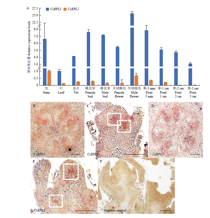
图3. CsRPL1/2表达模式分析
A:CsRPL1/2在黄瓜各器官中的表达分析;B—F:CsRPL1(B和C)和CsRPL2(D和E)在黄瓜果实与植株顶点处的原位杂交分析;B、D:CsRPL1/2在胎座框处表达;C、E:CsRPL1/2信号在植物顶端分生组织的CZ区域富集;白色虚线框代表信号富集区域;F:CsRPL-SP6探针作为负对照,未发现杂交信号。比例尺:100 μm
Fig. 3. Gene expression pattern of CsRPL1/2
A: Relative expression of CsRPL1/2 in various parts of cucumber; B-F: In situ hybridization analysis of CsRPL1 (B, C) and CsRPL2 (D, E) in cucumber fruits and shoot tips; B, D: CsRPL1/2 expressed in placenta C, E: CsRPL1/2 signal was enriched in the central zone of the meristem in the shoot tips; White dotted box represents the signal enrichment region; F: CsRPL-SP6 probe was used as negative control, and no hybridization signal was found. Scale bar: 100 μm