)
)
)
)
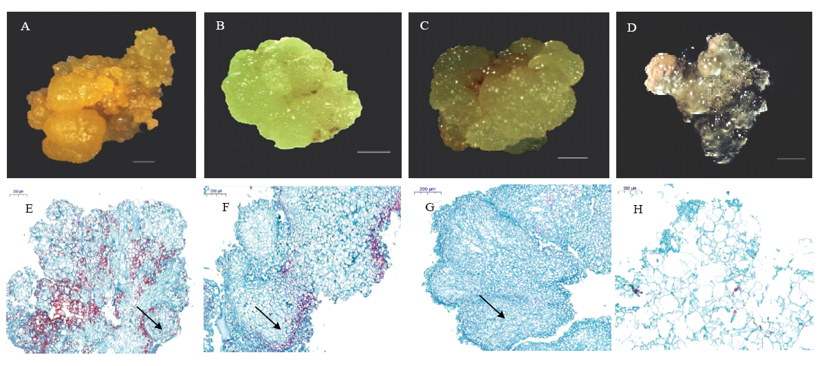
图5. ‘秋蜜红’不同状态愈伤组织形态学及细胞学观察
A:黄色疏松状胚性愈伤组织;B:绿色紧实状胚性愈伤组织;C:浅黄色透明状胚性愈伤组织;D:黄白色水渍状非胚性愈伤组织;E:黄色疏松状胚性愈伤组织细胞组织切片(箭头所示为球形胚结构);F:绿色紧实状胚性愈伤组织细胞组织切片(箭头所示为心形胚结构);G:浅黄色透明状胚性愈伤组织细胞组织切片(箭头所示为尚未完全形成的鱼雷形胚结构);H:黄白色水渍状非胚性愈伤组织细胞组织切片。A—D:图标比例尺为1 mm;E—H:图标比例尺为200 μm
Fig. 5. The morphological and cytological observation of different forms of calli from P. perscia ‘Qiumihong’
A: The yellow, loose embryogenic callus; B: The green, compact embryogenic callus; C: The pale yellow, transparent embryogenic callus; D: The yellow-white, dropsical non-embryonic callus; E: Microscope slide of the yellow, loose embryogenic callus (arrow indicated globular embryo); F: Microscope slide of the green, compact embryogenic callus (arrow indicated heart-shape embryo); G: Microscope slide of the pale yellow, transparent embryogenic callus (arrow indicated not yet fully formed torpedo-shape embryo); H: Microscope slide of the yellow-white, dropsical embryonic callus. A-D: Bar = 1 mm; E-H: Bar = 200 μm