p66Shc在绵羊卵母细胞中的表达及其与胞质氧化还原稳态的关系
张通1,2,栗瑞兰1,2,范晓梅1,3,刘春洁1,海日汗1,霍敏1,张家新1( )
)
)
Expression of p66Shc and Its Relationship with Cytoplasmic Redox Homeostasis in Sheep Oocytes
ZHANG Tong1,2,LI RuiLan1,2,FAN XiaoMei1,3,LIU ChunJie1,HAI RiHan1,HUO Min1,ZHANG JiaXin1()
)
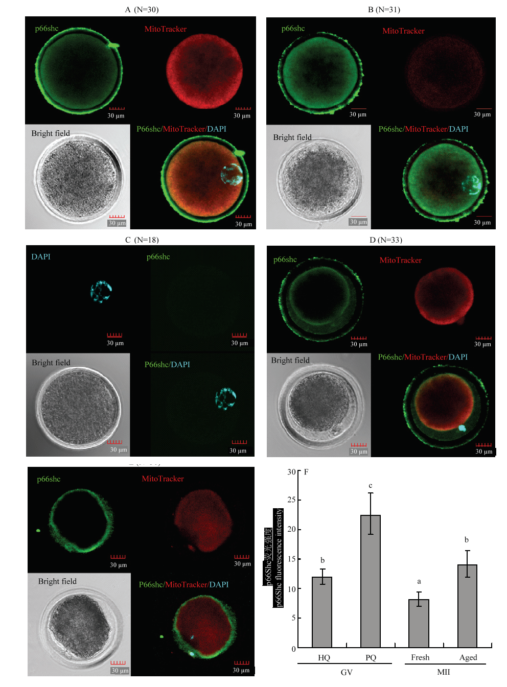
图3. p66Shc蛋白与线粒体在成熟前后不同质量卵母细胞的共定位
A:成熟前优质卵母细胞;B:成熟前劣质卵母细胞;C:阴性对照(成熟前);D:常规24 h成熟卵母细胞;E:成熟后老化的卵母细胞;F:p66Shc蛋白荧光强度。绿色标记p66Shc,红色标记线粒体,蓝色标记细胞核,比例尺表示30 μm。不同字母表示差异显著(P<0.05), 相同字母表示差异不显著(P>0.05)
Fig. 3. Co-localization of p66Shc protein and mitochondria in different quality oocytes before and after maturation
A: High quality immature oocytes; B: Poor quality immature oocytes; C: Negative control (immature oocytes); D: Conventional 24 h matured oocytes; E: Aged oocytes; F: p66Shc protein fluorescence intensity. Green labeled p66Shc, red labeled mitochondrial, blue labeled nuclear, the scale bar represents 30 μm. The different superscripts mean significant difference (P<0.05), and the same superscripts mean no significant difference (P>0.05)